
A microdevice and microscope system for studying egg cells
A microdevice and egg cell technology, which is applied in the field of microdevices and microscope systems for studying egg cells, can solve problems such as difficulty in measuring egg cell permeability parameters, and achieve the effect of a reliable test environment
- Summary
- Abstract
- Description
- Claims
- Application Information
AI Technical Summary
Problems solved by technology
Method used
Image

Examples
Embodiment
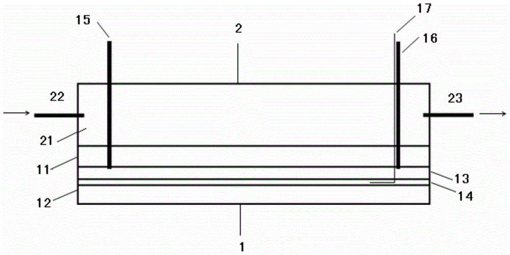
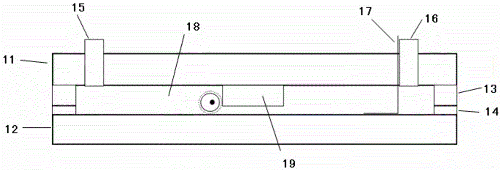
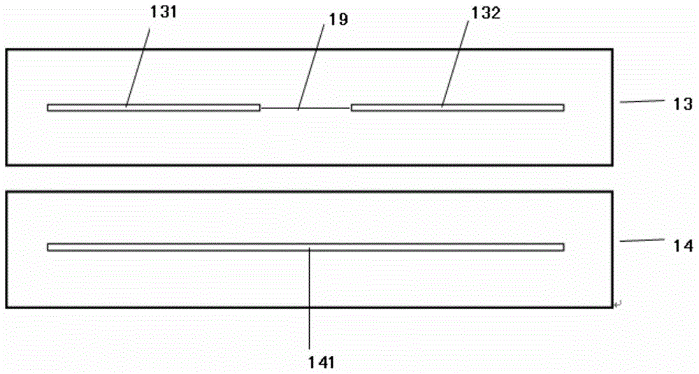
[0070] Structure of a microdevice used to study egg cells see Figure 1 to Figure 5 , wherein, the upper wall (the first cover) 11 and the lower wall (the second cover) 12 are silica glass; the first film 13 and the second film 14 are silica gel films, the thickness is 100 microns, 50 microns. The first film 13 is engraved with two short grooves 131 and 132 , and the second film 14 is engraved with a long groove (second groove) 141 . The width of the long groove and the two short grooves are both 200 μm, and the lengths are 44 mm, 30 mm, and 15 mm, respectively, and the outer edges are aligned. The first short groove 131 and the long groove 141 form a material chamber with a height of 150 microns, and the part between the two short grooves forms a baffle plate 19 with a height of 100 microns. The inlet conduit 15 is close to the edge of the material chamber, and the outlet conduit 16 is close to the other edge of the microperfusion chamber; both the inlet conduit 15 and the o...
PUM
| Property | Measurement | Unit |
|---|---|---|
| thickness | aaaaa | aaaaa |
| thickness | aaaaa | aaaaa |
| width | aaaaa | aaaaa |
Abstract
Description
Claims
Application Information