Stereotaxic frame for fusion of pet, ct and mr images of the abdomen and pelvis

An image fusion and stereotaxic technology, which is applied in the fields of application, medical science, X-ray/γ-ray/particle irradiation therapy, etc., can solve problems such as lack of body parts, achieve application value enhancement, simple installation and operation, improve efficiency and reliability effect

Active Publication Date: 2017-06-23
THE SECOND HOSPITAL OF TIANJIN MEDICAL UNIV
View PDF6 Cites 0 Cited by
  • Summary
  • Abstract
  • Description
  • Claims
  • Application Information

AI Technical Summary

Problems solved by technology

This method is widely used in the head and neck as a main registration fusion method, but there is no body (especially the abdomen and pelvis) stereotaxic framework

Method used


Image

  • Stereotaxic frame for fusion of pet, ct and mr images of the abdomen and pelvis
  • Stereotaxic frame for fusion of pet, ct and mr images of the abdomen and pelvis

Examples


Embodiment Construction

[0021] The technical solutions of the present invention will be further described in detail below in conjunction with the accompanying drawings and specific embodiments.

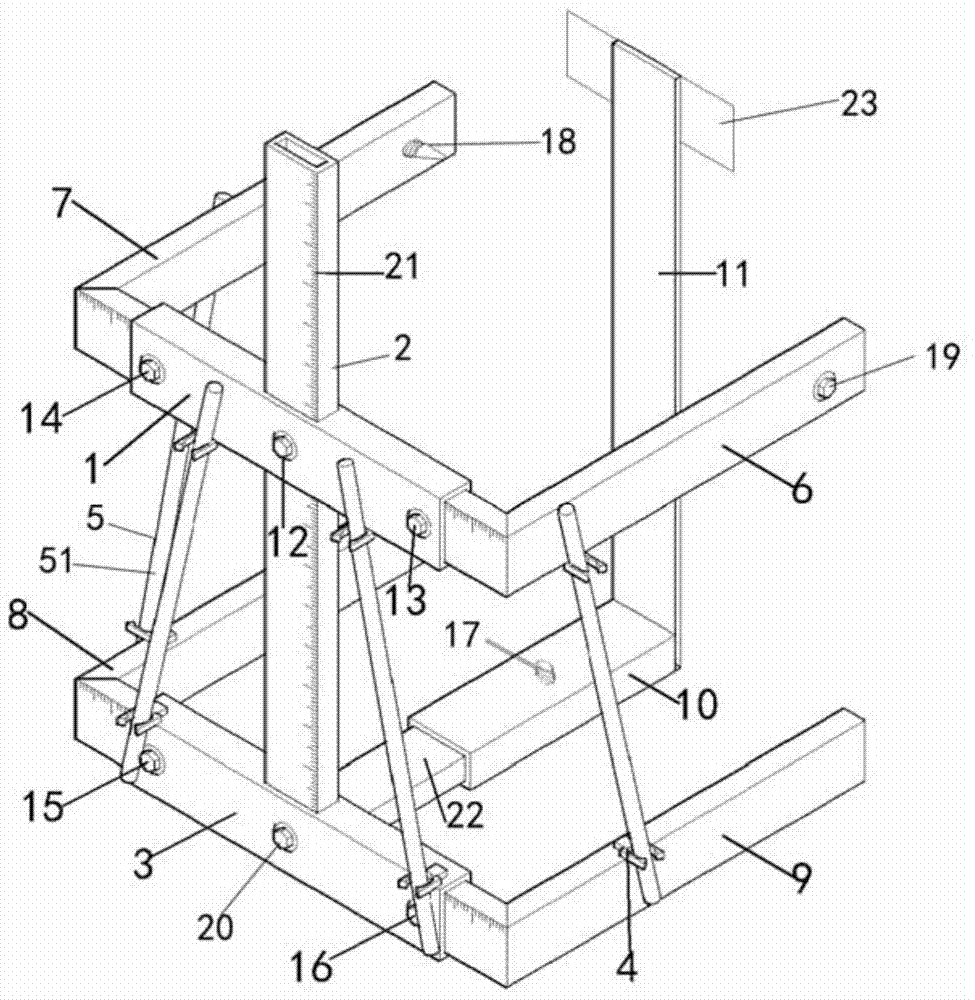
[0022] Such as figure 1 and figure 2 As shown, the present invention is a stereotaxic frame for fusion of PET, CT and MR images in the abdomen and pelvis, including a first bracket, a second bracket and a bracket.

[0023] The first bracket and the second bracket are U-shaped frames respectively, and the basic structure of the first bracket and the second bracket is the same, as figure 2 As shown, the first bracket consists of a sleeve 1 and right-angled plates 6, 7 inserted on both sides of the sleeve 1, and the second bracket consists of a sleeve 3 and right-angled plates 8, 9 inserted on both sides of the sleeve. constitute. In the first support and the second support, the opening width of the U-shaped frame is determined by adjusting the relative positions of the right-angle plates on both sides and...


PUM


Abstract

The invention discloses a stereotactic frame for fusion of PET, CT and MR images in the abdomen and pelvis, which includes U-shaped first and second brackets and brackets, and the opening width of the U-shaped frame is adjustable. The first bracket is parallel to the second bracket, the distance between the first bracket and the second bracket is adjustable, the bracket intersects the two brackets perpendicularly, a pointed top wire is provided on the first bracket, and the pointed top wire pushes from the outside of the U-shaped frame to the inside There are more than three positioning rods connected between the first and second brackets, at least two positioning rods intersect, and the positioning rods are detachable. The positioning rods include three sets of canned PET imaging agents and CT imaging agents respectively. and MR imaging agent positioning stick; the positioning stick adds positioning information for three different image data of PET, CT or MR. The invention is easy to operate, can be used for PET, CT or MR machines of any brand and model, realizes the main functions of expensive PET / CT, PET / MR machines, and improves the application value of individual equipment.

Description

technical field [0001] The invention relates to an auxiliary device for medical imaging diagnosis and treatment, in particular to a stereotaxic frame for the abdomen and pelvis. Background technique [0002] With the continuous advancement of modern medicine, radiology and computer technology, the development of medical imaging technology is changing with each passing day, and various medical imaging examination methods based on different imaging principles emerge in endlessly. So far, the imaging modes of medical imaging can be divided into two categories: anatomical structure imaging and functional metabolic imaging. Among them, CT (Computed Tomography) and MR (Nuclear Magnetic Resonance) can reflect the anatomical structure information of the human body and can perform three-dimensional imaging. CT has fast scanning speed, high spatial resolution, and clear imaging of rigid body (bone, joint, etc.) lesions, but the imaging of soft tissue (vascular, nerve, prostate, joint...

Claims


Application Information

Patent Timeline
Patent Type & Authority Patents(China)
IPC IPC(8): A61B6/03A61B5/055A61N5/10A61B90/00
Inventor 赵阳张雪宁牛远杰蒋宁贾荣荣彭景张恩龙李京津徐国萍
Owner THE SECOND HOSPITAL OF TIANJIN MEDICAL UNIV
Why Eureka
Try Eureka