X-ray CT-fluorescence imaging apparatus and method of single-source-emission and dual-mode imaging

A technology of fluorescence imaging and imaging method, applied in the field of X-ray CT-fluorescence imaging system, can solve the problems of low luminous efficiency and unstable light intensity, and achieve the effect of low coupling degree

Inactive Publication Date: 2016-04-27
SOUTHEAST UNIV
View PDF9 Cites 16 Cited by
  • Summary
  • Abstract
  • Description
  • Claims
  • Application Information

AI Technical Summary

Problems solved by technology

Most of the currently used fluorescent materials are autofluorescent, with low luminous efficiency and unstable light intensity.

Method used


Image

  • X-ray CT-fluorescence imaging apparatus and method of single-source-emission and dual-mode imaging
  • X-ray CT-fluorescence imaging apparatus and method of single-source-emission and dual-mode imaging

Examples


Embodiment Construction

[0045] The present invention will be further described below in conjunction with embodiment and accompanying drawing.

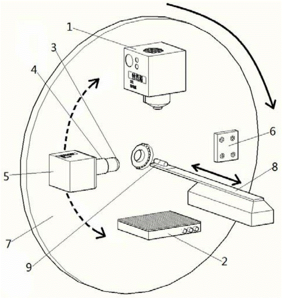
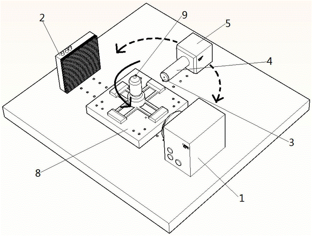
[0046] The invention provides an X-ray CT-fluorescence imaging system with single-source emission and dual-mode imaging. The system includes two structures. Structure A is characterized by using a turntable to drive the point source and two detectors to move together to obtain dual-mode images at various angles. Structure B obtains dual-mode images at various angles through the rotation of the stage. like.

[0047] The present invention designs an X-ray CT-fluorescence imaging system with single-source emission and dual-mode imaging. The system structure A involves a turntable, and designs an X-ray optical path system and a fluorescent optical path installed on the turntable. system. In the X-ray optical system, the cone beam rays are emitted by a point source, and after passing through the object under inspection, they are received by the X-ray detector to...


PUM


Abstract

The invention discloses an X-ray CT-fluorescence imaging apparatus and method of single-source-emission and dual-mode imaging. The method, by emitting X ray from a single source and exciting a fluorescent material, can achieve simultaneous dual-mode imaging through the X ray and a fluorescent detector. The apparatus comprises an X-ray light source, a carrying platform, an X-ray detector and the fluorescent detector which are arranged on the same plane, wherein the X-ray light source, the carrying platform and the X-ray detector are arranged on the same line, so that an X-CT light path is constituted; a fluorescent lens which is arranged in the fluorescent detector is arranged in a mode of aligning with the carrying platform; and the relative positions of the X-ray light source, the X-ray detector and the fluorescent detector are fixed, and the X-ray light source, the X-ray detector and the fluorescent detector can rotate relative to the carrying platform by taking the carrying platform as a center.

Description

technical field [0001] The invention belongs to the field of multi-mode molecular imaging, and relates to an X-ray CT-fluorescence imaging system for single-source emission and dual-mode imaging. Background technique [0002] Multimodal imaging systems play an important role in scientific research and medical testing, and have been widely used. From multimodal imaging equipment for small animals to human medical testing equipment, multimodal imaging has attracted widespread attention for its unique function of combining the advantages of different imaging equipment. Multimodal imaging systems currently mainly include: PET-CT system, ultrasound-CT / MR fusion imaging system, etc. [0003] PET-CT system is a multimodal imaging system widely used at present. It combines PET and CT systems and can effectively detect structural information and biological function information. However, the radiation dose of the PET-CT system is relatively large, and X-ray sources and radioactive...

Claims


Application Information

Patent Timeline
Patent Type & Authority Applications(China)
IPC IPC(8): A61B6/03A61B6/00
CPCA61B6/485A61B6/03A61B6/4208A61B6/5211
Inventor 顾宁沈涛孙翌李光罗守华陈功
Owner SOUTHEAST UNIV
Why Eureka
Try Eureka