Medical image processing device based on computer

A medical imaging and processing device technology, applied in the field of medical imaging, can solve the problems of inability to automatically rotate the angle, intelligent single image transmission, etc., and achieve the effect of simple structure and favorable production and processing.

Inactive Publication Date: 2017-08-11
南京译海网络科技股份有限公司
View PDF5 Cites 0 Cited by
  • Summary
  • Abstract
  • Description
  • Claims
  • Application Information

AI Technical Summary

Problems solved by technology

[0003] The intelligent single image transmission of the existing B-ultrasound probe can not automatically rotate the angle. The rotation of the angle

Method used


Image

  • Medical image processing device based on computer
  • Medical image processing device based on computer

Examples


Example Embodiment

[0015] In order to further understand the content, features, and effects of the present invention, the following embodiments are listed in conjunction with the accompanying drawings.

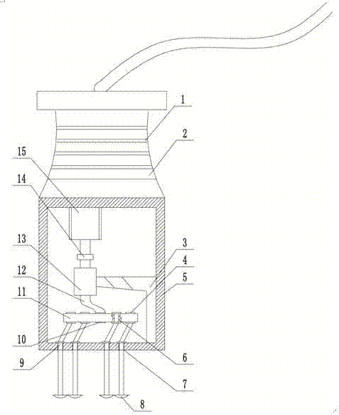
[0016] Combine below Figure 1-2 The structure of the computer-based medical image processing device of the present invention will be described in detail.

[0017] A computer-based medical image processing device includes a housing 4, the lower end of the housing 4 is provided with four through holes 7, and the inner wall of the housing 4 is fixedly provided with an L-shaped frame 3, the L-shaped frame 3 A ring sleeve 13 is provided at one end of the L-shaped frame 3, and the L-shaped frame 3 is a multi-segment detachable and connected soft structure, which can be specifically a metal strip coated with a colloid. When the area of ​​the ring sleeve 13 becomes larger, the L-shaped frame 3 can be used. Disassembly; the annular sleeve 13 is provided with a crankshaft 12, one end of the crankshaft 12 and...


PUM


Abstract

The invention belongs to the field of medical imaging, and relates to a medical image processing device based on a computer. The medical image processing device includes a shell, the lower end face of the shell is provided with four through holes, an L-shaped bracket is fixed to the side face of the inner wall of the shell, one end of the L-shaped bracket is provided with a circular sleeve, a first crank shaft is arranged in the circular sleeve, one end of the first crank shaft is connected with an output shaft of a motor through a coupler, the motor is fixed to the upper end face of the inner wall of the shell, the other end of the first crank shaft is rotationally connected with the center of a rotary table, four round holes are uniformly formed in the rotary table, second crank shafts are arranged in the round holes, the middles of the second crank shafts pass through the through holes, one end of each second crank shaft is provided with an image collection device, the other end of the first crank shaft is provided with a first limit protrusion, and the other end of each second crank shaft is provided with a second limit protrusion. The medical image processing device based on the computer can transmit multiple images and automatically rotate.

Description

technical field [0001] The invention belongs to the field of medical imaging, in particular to a computer-based medical image processing device. Background technique [0002] Imaging technology can comprehensively and accurately obtain various quantitative and qualitative data of patients, and provide correct digital information for diagnosis, treatment planning, operation and postoperative evaluation. Among them, ultrasound imaging technology plays an important role in all aspects of clinical medicine because of its low price, no damage to tissue, real-time and other advantages. B-mode ultrasound is a new subject, which has developed rapidly in recent years, and it has become an indispensable diagnostic method in modern clinical medicine. B-ultrasound can clearly display various cross-sectional images of various organs and surrounding organs. Because the images are full of solidity and close to the real structure of anatomy, the application of ultrasound can make an early ...

Claims


Application Information

Patent Timeline
IPC IPC(8): A61B8/00
CPCA61B8/44A61B8/48A61B8/52
Inventor 不公告发明人
Owner 南京译海网络科技股份有限公司
Why Eureka
Try Eureka