Three-dimensional ultrasound imaging method and device

A three-dimensional ultrasound and ultrasound echo technology, applied in ultrasonic/sonic/infrasonic diagnosis, sonic diagnosis, infrasonic diagnosis, etc., can solve problems such as difficulty in adjustment and lack of understanding of 3D space.

Active Publication Date: 2020-10-20
SHENZHEN MINDRAY BIO MEDICAL ELECTRONICS CO LTD
View PDF6 Cites 2 Cited by
  • Summary
  • Abstract
  • Description
  • Claims
  • Application Information

AI Technical Summary

Problems solved by technology

However, doctors need to have a very deep understanding of 3D space to be able to adjust the median sagittal section through manual rotation and translation geometric operations in 3D. Difficult to manually adjust the midsagittal plane from individual data

Method used


Image

  • Three-dimensional ultrasound imaging method and device
  • Three-dimensional ultrasound imaging method and device
  • Three-dimensional ultrasound imaging method and device

Examples


Embodiment 1

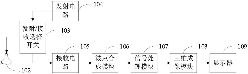
[0038] This embodiment provides a three-dimensional ultrasonic imaging device, its structural block diagram is as follows figure 1 shown. The three-dimensional ultrasonic imaging device includes a probe 102 , a transmit / receive selection switch 103 , a transmit circuit 104 , a receive circuit 105 , a beam forming module 106 , a signal processing module 107 , a three-dimensional imaging module 108 and a display 109 . The transmitting circuit 104 sends a group of pulses with delayed focus to the probe 102, and the probe 102 transmits ultrasonic waves to the body tissue under test (not shown in the figure), and receives the tissue-bearing tissue reflected from the body tissue under test after a certain delay. The ultrasonic echo of the information is converted back into an electrical signal. The receiving circuit 105 receives the ultrasonic echo signals converted into electrical signals, and sends the ultrasonic echo signals to the beamforming module 106 . The ultrasonic echo s...

Embodiment 2

[0069] The three-dimensional ultrasound imaging method / system provided in this embodiment is similar to Embodiment 1, and the same parts of the two will not be repeated here. The difference is that in step 25 of Embodiment 1, the fetus in the mid-sagittal section is determined by the orientation of the skull. and the step 25 of this embodiment is to determine the head orientation of the fetus in the midsagittal section according to the orientation of the transparent compartment.

[0070] Such as Figure 5 and Figure 8 As shown, in the ultrasound image, the shape of the transparent septum is a curved crescent. When the fetus is upright in the ultrasound image, the transparent septum appears as an upward crescent shape. On the contrary, when the fetus is inverted in the ultrasound image , the hyaline compartment exhibits a downwardly convex crescent shape. Therefore, the orientation of the fetal head on the midsagittal plane can be judged according to the orientation of the l...

Embodiment 3

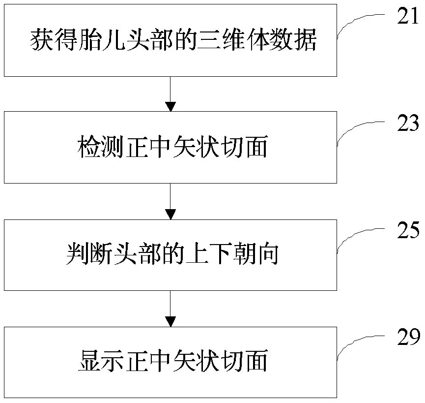
[0080] The three-dimensional ultrasonic imaging method provided in this embodiment is as follows: Figure 12 As shown, it includes: step 21 of obtaining the three-dimensional volume data of the fetal head, step 23 of detecting the midsagittal section, step 27 of judging the face orientation, and step 29 of displaying the midsagittal section. Wherein, steps 21, 23 and 29 are the same as those in embodiment 1 or 2, and will not be repeated here. Based on the method, this embodiment also provides a three-dimensional ultrasonic imaging device for implementing the method. For the structure of the device except for the three-dimensional ultrasonic imaging part, reference may be made to the foregoing embodiment 1, which will not be repeated here.

[0081] Because the transparent compartment is located at the front of the cranium, the face and back of the fetus (that is, the face orientation of the fetus) can be determined through the position of the transparent compartment.

[0082]...


PUM


Abstract

A three-dimensional ultrasonic imaging method and a three-dimensional ultrasonic imaging device. The three-dimensional ultrasonic imaging method includes the following steps: transmitting ultrasonic waves to the fetal head; receiving ultrasonic echoes to obtain ultrasonic echo signals; obtaining a three-dimensional volume of the fetal head according to the ultrasonic echo signals. Data; according to the characteristics of the mid-sagittal section of the fetal head, the mid-sagittal section is detected from the three-dimensional volume data; the orientation of the fetal head in the mid-sagittal section is determined; according to the orientation of the fetal head, the mid-sagittal section Displayed as an image suitable for observation. The mid-sagittal section image is processed and displayed as an image conforming to human observation habits, which makes it easier for doctors to identify and observe the situation of the mid-sagittal section of the fetal brain.

Description

technical field [0001] The invention relates to the technical field of medical ultrasonic imaging, in particular to a three-dimensional ultrasonic imaging method and device. Background technique [0002] Ultrasound instruments are generally used by doctors to observe the internal tissue structure of the human body. The doctor places the ultrasonic probe on the skin surface corresponding to the part of the human body to obtain an ultrasonic image of the part. Ultrasound has become one of the main auxiliary methods for doctors' diagnosis due to its safety, convenience, non-destructive, and low-cost characteristics. Among them, obstetrics is one of the most widely used fields of ultrasound diagnosis. In this field, ultrasound avoids the influence of X-rays on the mother and fetus, and its application value is obviously superior to other imaging examination equipment. Ultrasound can not only observe and measure fetal morphology, but also obtain a variety of physiological and pa...

Claims


Application Information

Patent Timeline
Patent Type & Authority Patents(China)
IPC IPC(8): A61B8/14
CPCA61B8/0808A61B8/4405A61B8/4427A61B8/4472A61B8/466A61B8/483A61B8/0866A61B8/5223A61B8/523G16H50/30A61B8/14A61B8/0816
Inventor 邹耀贤林穆清陈志杰
Owner SHENZHEN MINDRAY BIO MEDICAL ELECTRONICS CO LTD
Why Eureka
Try Eureka