Mitral valve opening area detection method, system and device based on artificial intelligence

A technology of artificial intelligence and detection methods, applied in the field of detection and recognition of medical video images, can solve problems such as large differences between individuals, and achieve the effect of improving accuracy and consistency

Active Publication Date: 2020-06-12
GENERAL HOSPITAL OF PLA
View PDF4 Cites 4 Cited by
  • Summary
  • Abstract
  • Description
  • Claims
  • Application Information

AI Technical Summary

Problems solved by technology

[0003] In the prior art, echocardiography is often used to measure the mitral valve orifice area, but since the results of echocardiography are mostly dependent on the experience of the examiner, there are large inter-individual differences

Method used


Image

  • Mitral valve opening area detection method, system and device based on artificial intelligence
  • Mitral valve opening area detection method, system and device based on artificial intelligence
  • Mitral valve opening area detection method, system and device based on artificial intelligence

Examples


Embodiment Construction

[0042] The application will be further described in detail below in conjunction with the accompanying drawings and embodiments. It should be understood that the specific embodiments described here are only used to explain related inventions, rather than to limit the invention. It should also be noted that, for ease of description, only parts related to the invention are shown in the drawings.

[0043] It should be noted that, in the case of no conflict, the embodiments in the present application and the features in the embodiments can be combined with each other. The present application will be described in detail below with reference to the accompanying drawings and embodiments.

[0044] Echocardiography, as described herein, is an ultrasound image that uses the special physical properties of ultrasound to examine the anatomy and functional status of the heart and great vessels.

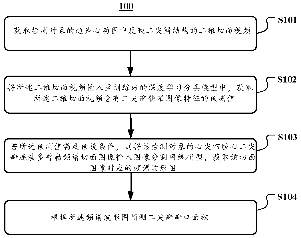
[0045] refer to figure 1 , which shows a method 100 for ultrasonic detection and recognition of...


PUM


Abstract

The invention discloses a mitral valve opening area detection method, system and device based on artificial intelligence. The method comprises the steps that a two-dimensional section video reflectinga mitral valve structure in an ultrasonic cardiogram of a detection object is acquired; inputting the two-dimensional section video into a trained deep learning classification model, and obtaining aprediction value of the two-dimensional section video containing mitral valve stenosis image features; if the prediction value meets a preset condition, inputting the heart tip four-cavity heart mitral valve continuous Doppler frequency spectrum section image of the detection object into an image segmentation network model, and obtaining a frequency spectrum oscillogram corresponding to the section image; and predicting the mitral valve opening area according to the frequency spectrum oscillogram. According to the method, the system and the equipment, the accuracy and the consistency of ultrasonic detection are greatly improved.

Description

technical field [0001] The present application relates to the technical field of detection and recognition of medical video images, in particular to an artificial intelligence-based mitral valve area detection method, system and equipment. Background technique [0002] Severe mitral stenosis (mitral stenosis, MS) will lead to a significant increase in left atrial pressure and left atrial enlargement, causing atrial fibrillation, hemoptysis, heart failure and other diseases. hospitalization, and even death. Mitral valve area is one of several indicators for assessing the severity of mitral stenosis and making surgical decisions. [0003] In the prior art, echocardiography is often used to measure the area of ​​the mitral valve orifice, but since the results of echocardiography mostly depend on the experience of the examiner, there are relatively large inter-individual differences. In recent years, studies have shown that artificial intelligence technology has great potentia...

Claims


Application Information

Patent Timeline
IPC IPC(8): G06T7/62A61B5/107
CPCA61B5/1075A61B5/1076G06T7/62G06T2207/10016G06T2207/10132G06T2207/20081G06T2207/20084G06T2207/30048G06T2207/30104
Inventor 李宗任何昆仑杨菲菲刘博罕王秋霜陈煦郭华源张璐邓玉娇
Owner GENERAL HOSPITAL OF PLA
Why Eureka
Try Eureka