Medical imaging device, method for supporting medical personnel, computer program product, and computer-readable storage medium

a medical imaging and medical technology, applied in the field of medical imaging devices and a method for supporting medical personnel, can solve the problems that tissue regions of the same organ cannot be readily distinguished or delineated from one another with conventional methods, and may only be insufficiently defined with markers, so as to achieve more flexible and reliable, less physical restrictions or impediments, and simple and robust tracking.

Active Publication Date: 2022-03-22
SIEMENS HEALTHCARE GMBH
View PDF9 Cites 1 Cited by
  • Summary
  • Abstract
  • Description
  • Claims
  • Application Information

AI Technical Summary

Benefits of technology

[0012]The data processing facility of the medical imaging device serves, and thus is designed and configured for the automatic registration of the image data with the 3D data record by alignment of the at least one detected tissue boundary on a corresponding segment or tissue boundary in a 3D data record. The image data and the 3D data record may thus be virtually overlaid or brought into coincidence, so that spatial points or regions corresponding to one another, in the present case in particular, the tissue boundary, are brought into coincidence with one another or into a spatial positional relationship to one another corresponding to reality or are assigned to one another accordingly. A corresponding assignment may be given by determining a transformation of corresponding coordinate systems of the 3D data record and of the image data into one another, so that by the determined transformation, the image data may be spatially correctly mapped onto the 3D data record or vice versa.
[0047]Equally, the tissue boundary or the tissue region delimited by the tissue boundary may be gathered from the image data, for example, by a corresponding, in particular automatic, segmentation and overlaid on the 3D data record. Equally, the overlay image may be an overlaying of the different individual images of the image data so that, for example, a fluorescence image which images or represents a fluorescence of one of the tissue regions in a non-visible spectral region and an optical image which represents or images the tissue regions in the optical spectral region are overlaid on one another to generate the overlay image. Therein optionally, additional virtual models or elements, for example, the 3D model generated from the 3D data record, may be inserted or overlaid. The overlay image may thus be, in particular, a mixture or a combination of three-dimensional and two-dimensional images or image contents. An overlay image of this type may advantageously offer a particularly comprehensive and content-rich support to the medical personnel.

Problems solved by technology

Such tissue regions of the same organ cannot be readily distinguished or delineated from one another with conventional methods and may only be insufficiently defined with markers.

Method used


Image

  • Medical imaging device, method for supporting medical personnel, computer program product, and computer-readable storage medium
  • Medical imaging device, method for supporting medical personnel, computer program product, and computer-readable storage medium
  • Medical imaging device, method for supporting medical personnel, computer program product, and computer-readable storage medium

Examples


Embodiment Construction

[0062]The components of the embodiments as described in the exemplary embodiments each represent individual features of the disclosure that are to be regarded as independent of one another and each also further develop the disclosure independently of one another and are thus also to be considered individually, or in a different combination from that shown, as a constituent part of the disclosure. Furthermore, the embodiments described are also enhanceable through others of the previously described features of the disclosure.

[0063]In the figures, elements which are identical, have the same function or correspond to one another are each provided with the same reference signs for the sake of clarity, even though they may represent different instances or examples of the corresponding elements.

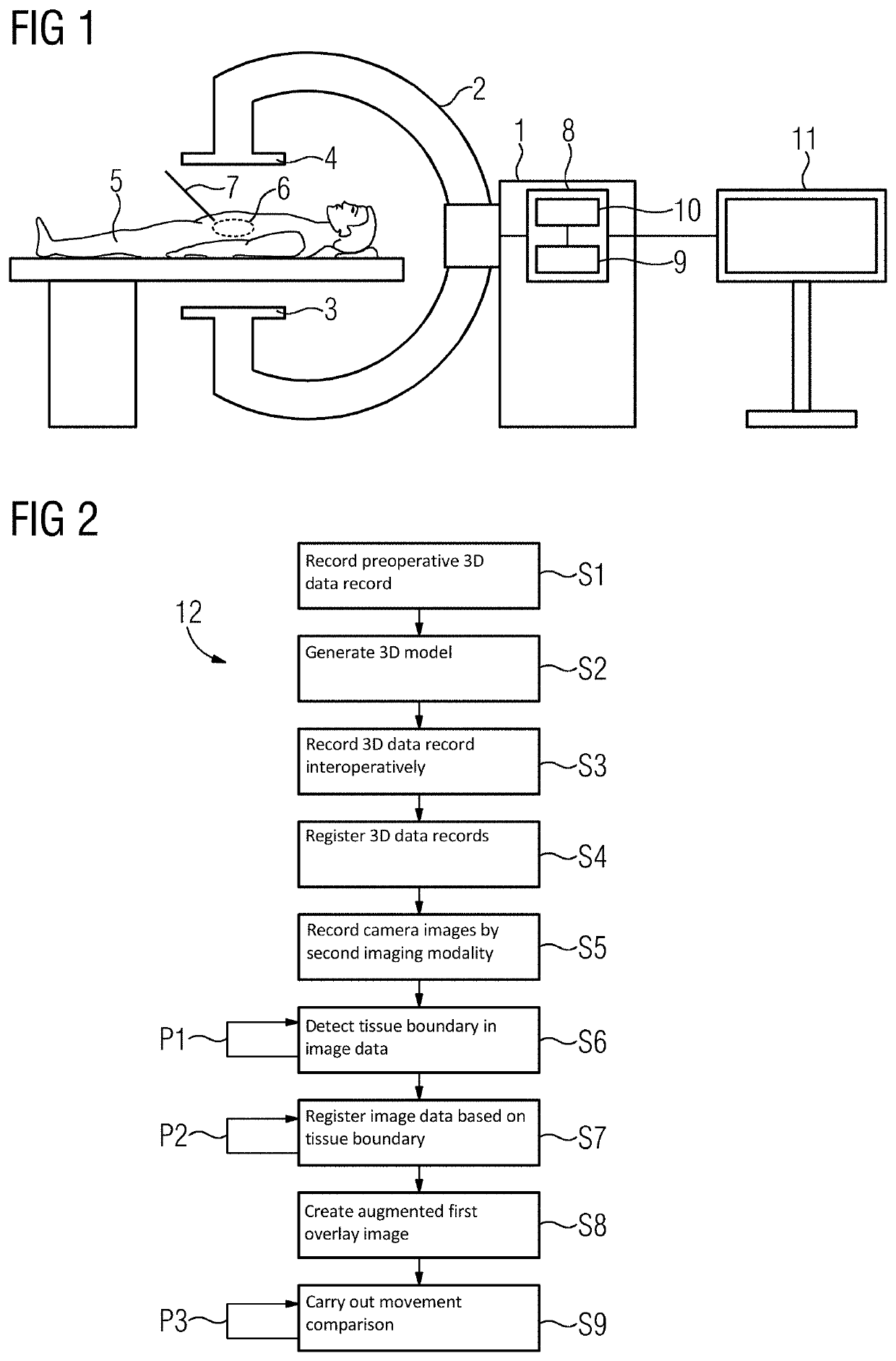
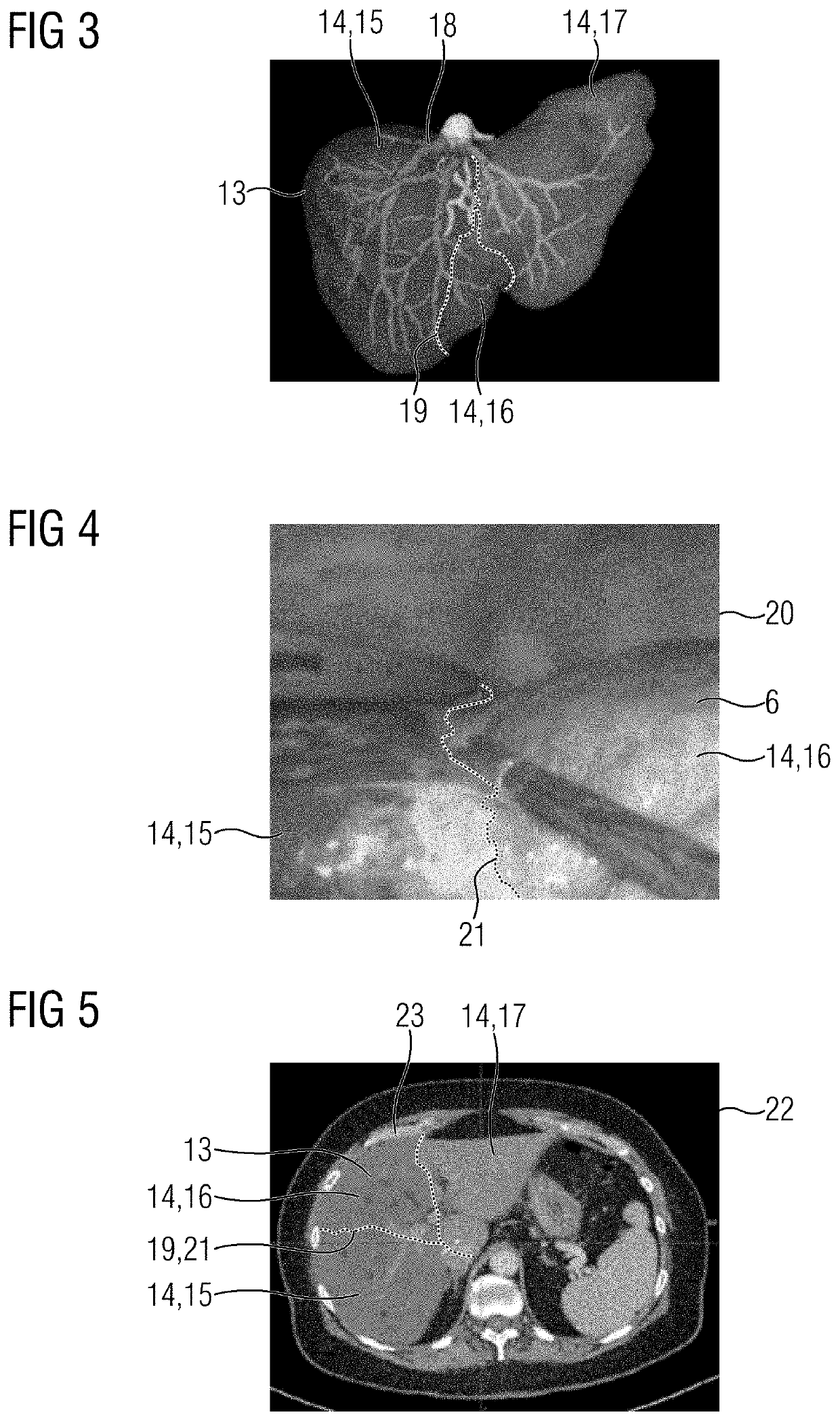
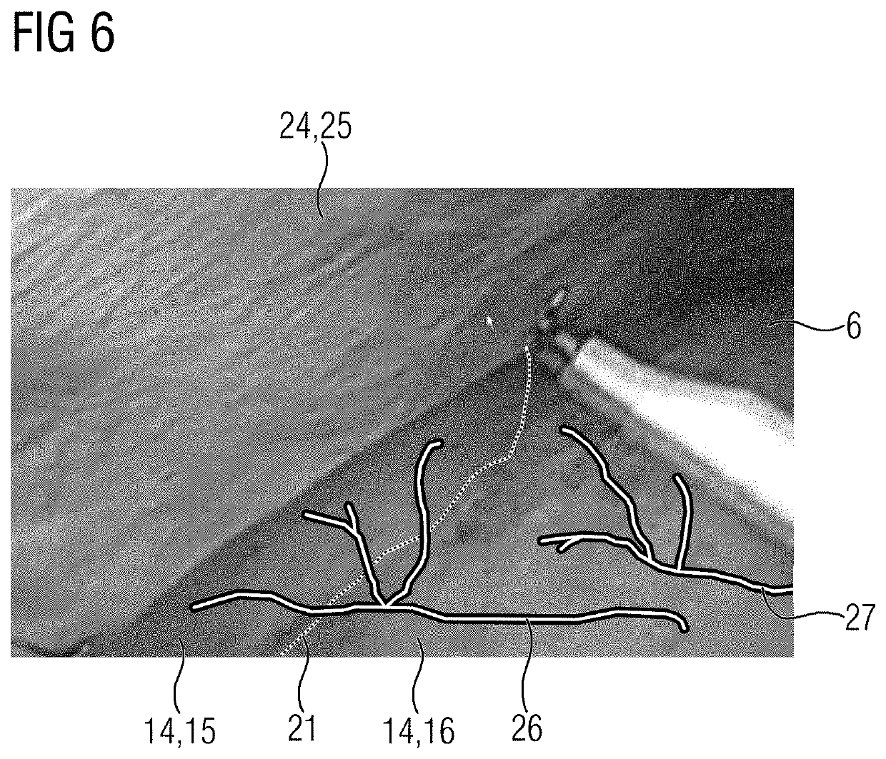
[0064]In the field of medical imaging technology, an image-based guidance and navigation is desirable for many applications, for example, for a liver resection, in particular if individual segments...


PUM


Abstract

The disclosure relates to a medical imaging device, a method for supporting medical personnel, a corresponding computer program product, and a corresponding computer-readable storage medium. In the method, a 3D data record is received from a first imaging modality and image data of an examination object is received from a second imaging modality. Different tissue regions of the same organ are imaged with different imaging properties at least under the second imaging modality. A tissue boundary is detected between the tissue regions in the image data. For registration of the image data with the 3D data record, the tissue boundary is aligned at a corresponding tissue boundary in the 3D data record. In addition, or alternatively, based on the tissue boundary, an automatic movement compensation takes place of a movement of the examination object which has occurred during the generation of the image data.

Description

[0001]The present patent document claims the benefit of German Patent Application No. 10 2019 200 786.4, filed Jan. 23, 2019, which is hereby incorporated by reference.TECHNICAL FIELD[0002]The disclosure relates to a medical imaging device and a method for supporting medical personnel. The disclosure further relates to a corresponding computer program product and a computer-readable storage medium on which a corresponding computer program product is stored.BACKGROUND[0003]In medical practice, it may be decisive for the success of a diagnosis and treatment that relevant medical personnel, for example, a physician or surgeon administering treatment, have the most precise and complete possible image of a relevant anatomical situation at the right time point. For this purpose, a whole range of measures and methods which may provide support to the medical personnel, for example, with a visualization have been developed in recent years. With conventional methods known from medical practic...

Claims


Application Information

Patent Timeline
Patent Type & Authority Patents(United States)
IPC IPC(8): G06K9/00G06T7/174G06T19/20G06T7/70
CPCG06T7/174G06T7/70G06T19/20G06T2207/10064G06T2207/30244G06T2219/2004A61B6/032A61B6/4417A61B5/055A61B6/466A61B6/463A61B6/5247A61B6/52A61B6/469G06T2200/04G06T2207/10081G06T2207/10088G06T2207/10012G06T2207/10132G06T2207/30056G06T2207/10024G06T7/33
Inventor REGENSBURGER, ALOISALZAGA, AMILCAR
Owner SIEMENS HEALTHCARE GMBH
Why Eureka
Try Eureka