Method and apparatus for aligning microscope images

a microscope and image technology, applied in the field of methods and equipment for aligning microscope images, can solve the problem of less easy determination of tissue structur

Active Publication Date: 2007-10-04
ROOM 4 GRP
View PDF7 Cites 4 Cited by
  • Summary
  • Abstract
  • Description
  • Claims
  • Application Information

AI Technical Summary

Problems solved by technology

In examining biological specimens, it is well known that there are problems.
On the other hand the tissue structure is less easily determined.
The difficulty is that it is necessary for an observer such as a pathologist to be

Method used


Image

  • Method and apparatus for aligning microscope images
  • Method and apparatus for aligning microscope images
  • Method and apparatus for aligning microscope images

Examples


Embodiment Construction

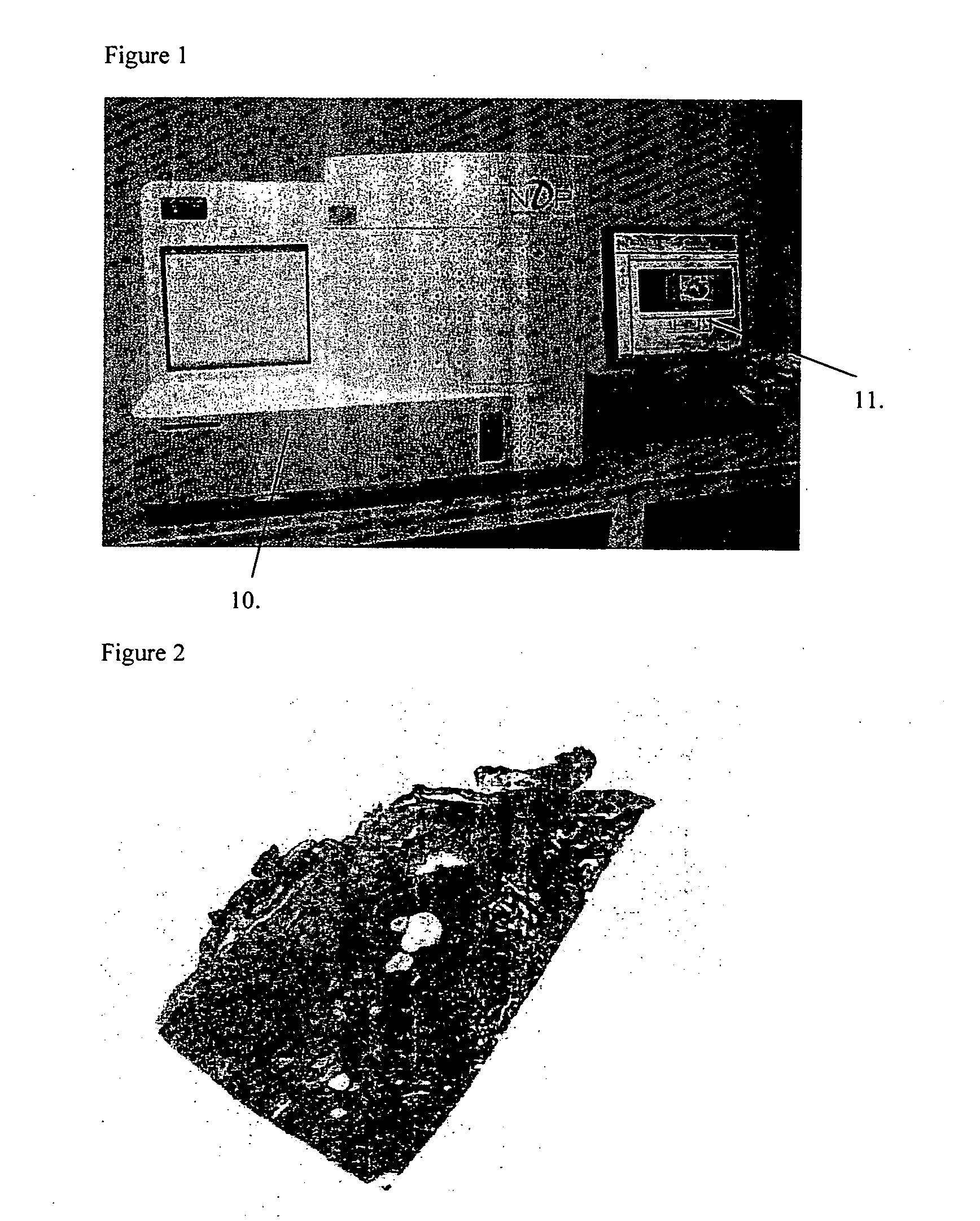
[0051] Referring to FIG. 1, there is shown a VM microscope 10 which comprises microscope optical components, a stage for mounting a specimen to be examined, a CCD (charge coupled device) array or other electronic imaging device for receiving the image of the specimen, a memory for storing the image, a computer for processing the image and a VDU 11 for displaying the image and other data. A prepared slide of, normally, a slice of biological tissue is scanned by the VM microscope to produce an image of the type shown in FIG. 2. For each point on the image on the CCD, the CCD array includes a red, green and blue wavelength detector providing red, green and blue signals. The operation of such a virtual microscope 10 is well known in the art.

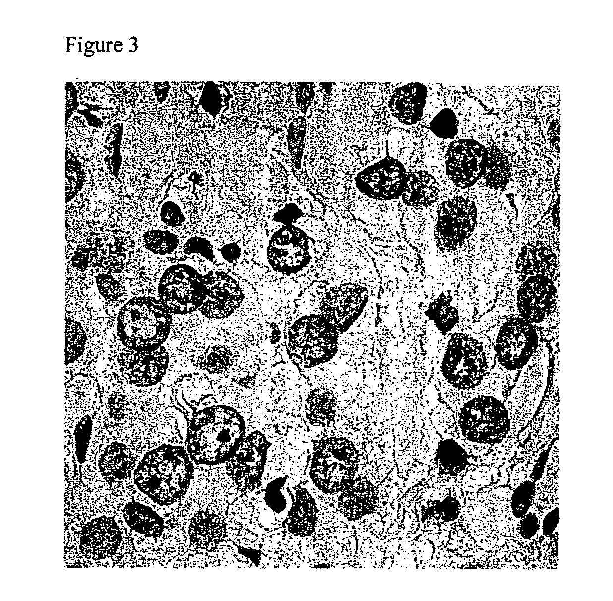
[0052] The image shown in FIG. 2 is of biological tissue which has been stained with a conventional H and E stain. This stain highlights the structure of the cellular material in the biological specimen.

[0053] In use, such an image may be examined ...


PUM


Abstract

A method and apparatus for aligning microscope images. Microscope images of the same or very similar subjects provided by different microscopes are aligned. The images from two types of microscope, such as a Virtual Microscope (VM) and a Light Microscope (LM) are used. An image produced by the virtual microscope is easily read by a viewer, as it represents a scan of a whole slide rather than individual high power fields of view. An area of the image can be selected for further examination or objective analysis by the LM microscope. The qualitative or quantitative information obtained from the Light microscope using the method described may then be located back into the virtual microscope image to provide understandable context.

Description

BACKGROUND TO THE INVENTION [0001] The present invention relates to a method and apparatus for aligning microscope images. The preferred arrangement of the invention relates to an arrangement in which microscope images of the same or very similar subjects provided by different microscopes or the same microscope at different magnifications are to be aligned. [0002] For example, microscope images of the same (or very similar) subject but at different powers, or different types of microscope image, for example, the same or similar microscope images stained with different types of stain, need to be compared and to compare the two images it is necessary to be able to align the two images or at least the parts of the image under examination. [0003] In examining biological specimens, it is well known that there are problems. The microscope slide of a biological specimen may be examined for the tissue structure which is particularly useful for a pathologist, using H and E staining (see http...

Claims


Application Information

Patent Timeline
IPC IPC(8): G06K9/32
CPCG02B21/367A61B5/0059G02B21/18
Inventor MADDISON, JOHNDANIELSEN, HAVARD E.
Owner ROOM 4 GRP
Why Eureka
Try Eureka