Optical coherence tomography imaging device for imaging a retina of a human subject

a coherence tomography and imaging device technology, applied in optics, medical science, instruments, etc., can solve the problems of visual dysfunction, scarring of the pigment epithelium, deterioration of acuity, etc., and achieve the effect of convenient use, compactness and low cos

Inactive Publication Date: 2017-06-22
MIMOSA
View PDF4 Cites 13 Cited by
  • Summary
  • Abstract
  • Description
  • Claims
  • Application Information

AI Technical Summary

Benefits of technology

[0007]It is the object of the invention to create an OCT imaging device pertaining to the technical field initially mentioned, that is inexpensive, compact and easy to use.

Problems solved by technology

An increase in vascular permeability leads to abnormal fluid collection within or below the retina that causes visual dysfunction when it involves the center of the macula.
This leads to rapidly deteriorating acuity, scarring of the pigment epithelium, and permanent visual loss or blindness.
For the OCT acquisition usually sophisticated and correspondingly expensive state-of-the-art OCT devices are employed.
This puts a considerable burden upon the patients.
However, today such devices are not available.

Method used


Image

  • Optical coherence tomography imaging device for imaging a retina of a human subject
  • Optical coherence tomography imaging device for imaging a retina of a human subject
  • Optical coherence tomography imaging device for imaging a retina of a human subject

Examples


Embodiment Construction

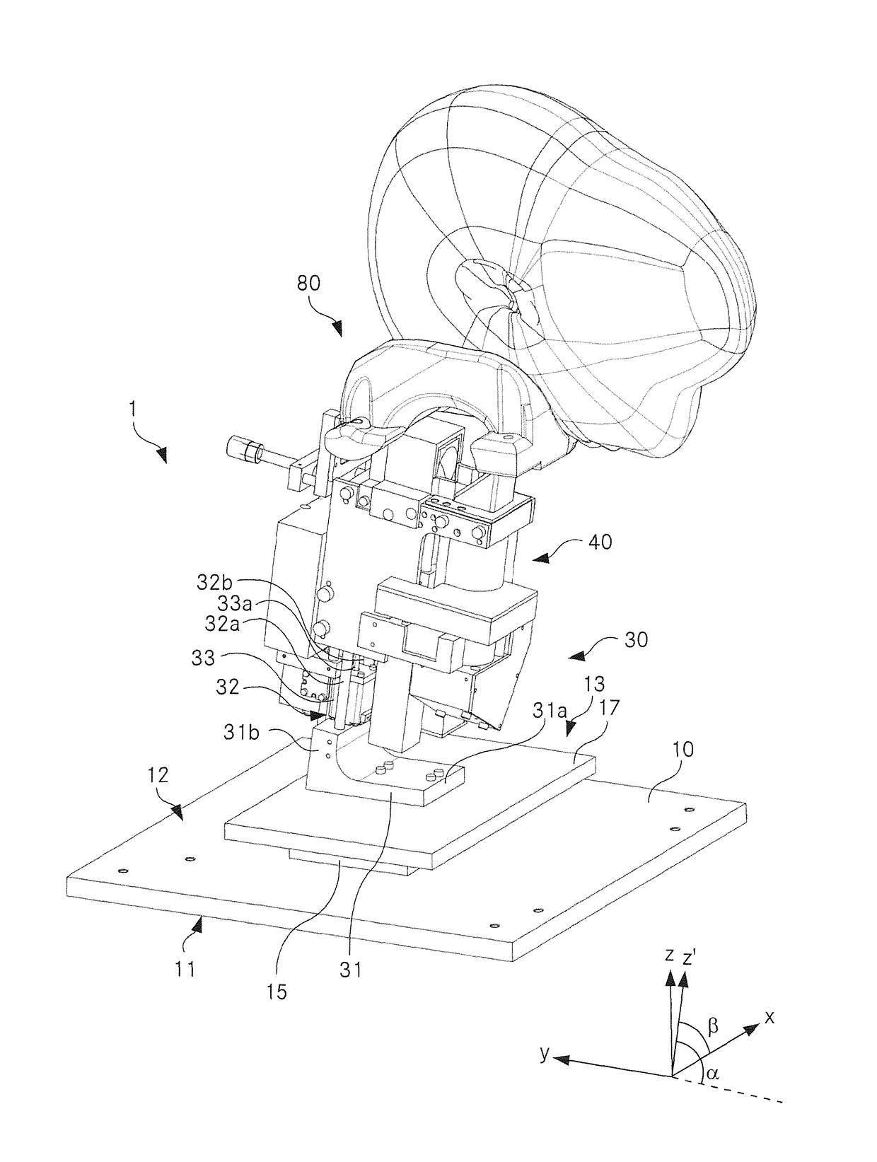
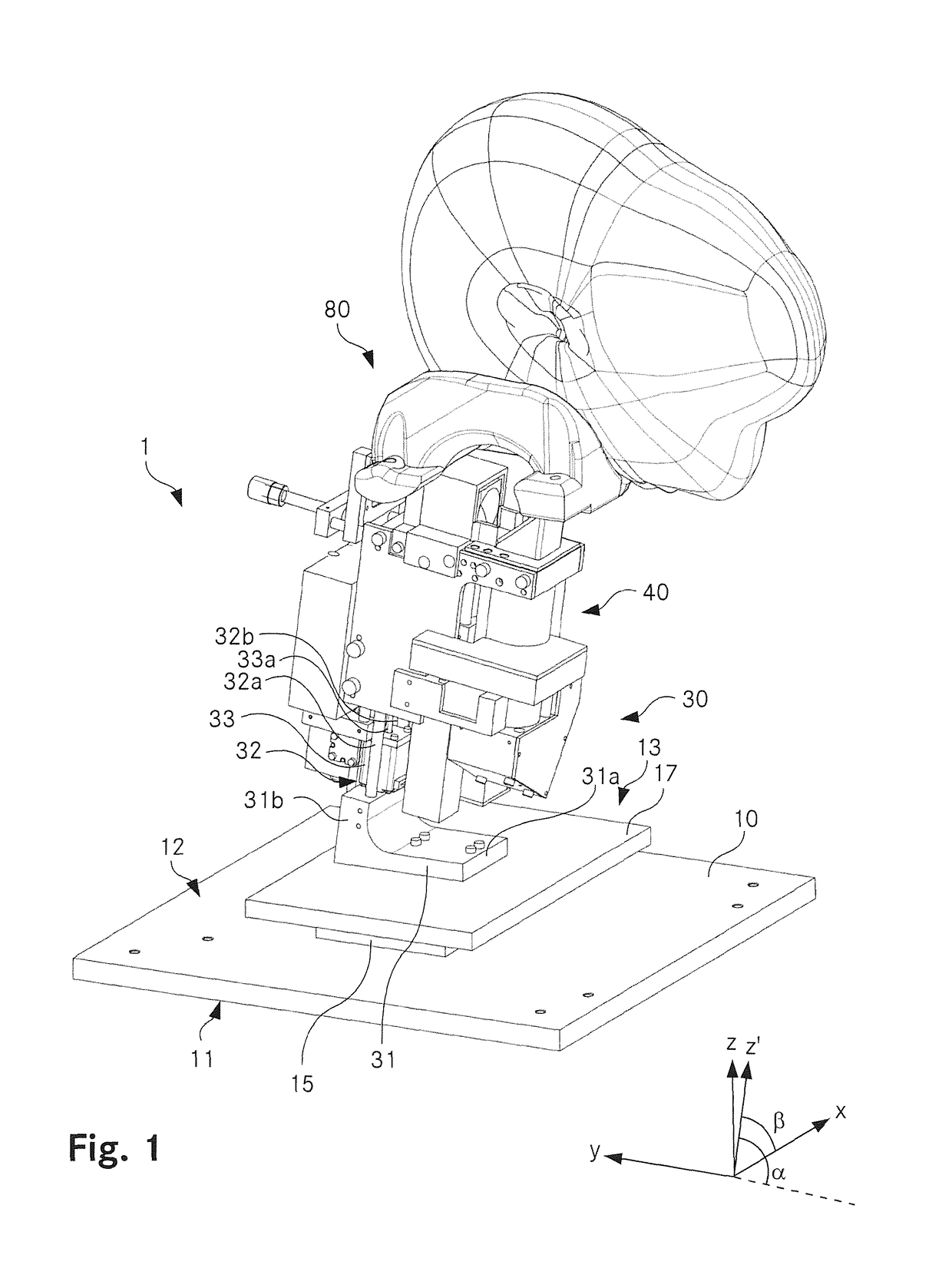
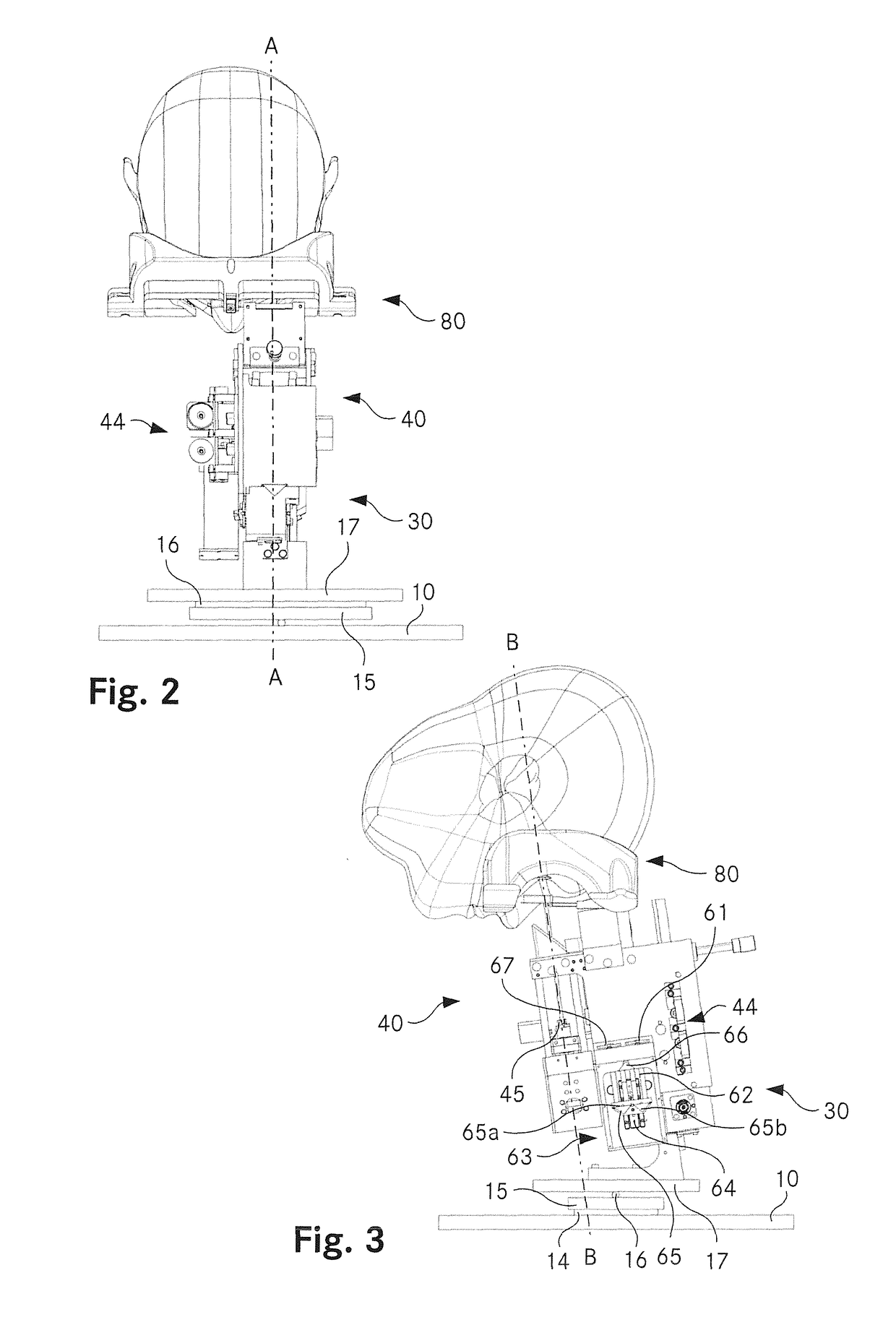
[0046]FIG. 1 shows an articulated view of an OCT device according to an embodiment of the invention. FIG. 2 shows a frontal view, FIG. 3 a side view of the OCT device, as seen from the right hand side. FIGS. 4 and 5 show cross-sectional views of the OCT device, FIG. 4 in a yz plane A-A shown in FIG. 2, FIG. 5 in the xz′ plane B-B shown in FIG. 3, seen from behind. For simplicity and in order to provide an overview, a housing surrounding the main optical unit as well as a spectrometer have been omitted in the Figures.

[0047]The main components of the OCT device 1 are a base plate 10, an optical unit 30 movable mounted to an upper surface of the base plate 10 and a head support 80 arranged above the optical unit 30.

[0048]The base plate 10 is rectangular and has uniform thickness. Its size is about 40×40 cm. The base plate 10 comprises a lower surface 11, which is a support surface for the OCT device 1 to be put on a flat surface such as a tabletop, and an upper surface 12 to which the ...


PUM


Abstract

An optical coherence tomography imaging device (1) for imaging a retina of a human subject comprises a base (10) having a planar contact surface (11), an optical unit (30) providing a sample beam to scan the retina of the human subject and a head support (80) to be contacted by a head portion of the human subject, the head support (80) defining an entrance position of the sample beam entering an eye of the human subject. An angle between the sample beam at the entrance position and the planar contact surface (11) of the base (10) is 50 to 90°. This arrangement ensures that the head rests in a stable and well defined position with respect to the OCT optics. The risk of negative effects on the imaging process due to head movements, in particular due to movements during the scan time of the OCT device is considerably reduced. Furthermore, the device may assume a compact form factor.

Description

TECHNICAL FIELD[0001]The invention relates to an optical coherence tomography imaging device for imaging a retina of a human subject, comprising a base having a planar contact surface, an optical unit providing a sample beam to scan the retina of the human subject and a head support to be contacted by a head portion of the human subject, the head support defining an entrance position of the sample beam entering an eye of the human subject.BACKGROUND ART[0002]Age-related Macular Degeneration (AMD) and especially neovascular AMD (nAMD) is the leading cause of blindness in the developed countries in people ageing over 50 years. An increase in vascular permeability leads to abnormal fluid collection within or below the retina that causes visual dysfunction when it involves the center of the macula. This leads to rapidly deteriorating acuity, scarring of the pigment epithelium, and permanent visual loss or blindness.[0003]However, intravitreal injection of antiangiogenic agents, includin...

Claims


Application Information

Patent Timeline
Patent Type & Authority Applications(United States)
IPC IPC(8): A61B3/10G02B27/00A61B3/00A61B3/14A61B3/12
CPCA61B3/102A61B3/14A61B3/1225G02B27/30A61B3/0075G02B27/0068A61B3/0083
Inventor KOWAL, JENSMALOCA, PETERRIEDER, BRUNO
Owner MIMOSA
Why Eureka
Try Eureka