3-dimensional model creation using whole eye finite element modeling of human ocular structures

a technology of ocular structure and 3D model, applied in the field of 3D model creation of complete ocular fem of human ocular accommodation, can solve the problems of inability to effectively restore the ability of presbyopia, lack of near vision focusing ability, and inability to develop effective treatments for presbyopia. to achieve the effect of restoring the ability

Inactive Publication Date: 2018-02-22
ACE VISION GRP INC
View PDF1 Cites 6 Cited by
  • Summary
  • Abstract
  • Description
  • Claims
  • Application Information

AI Technical Summary

Benefits of technology

[0017]Systems, methods and devices disclosed herein can be used to perform other functions as well, such as those pertaining to modelling other structures of the eye, such as the back of the eye, including: lamina cribrosa, Ocular Nerve Head and others, related to ocular structures and functions. For example, regarding the posterior globe: new insights and understanding of the lamina cribrosa are possible, as are insights into the complex structure of the peripapillary sclera, and attachments of the choroid using complex math for solving elastic and viscoelastic equations and simulations may provide additional benefits.

Problems solved by technology

As people age, they develop presbyopia and lose accommodative ability, leaving people over the age of 50 with an almost complete lack of focusing ability for near vision.
Although scientists have studied accommodation for centuries the functional mechanism is not well understood.
Without understanding the interactions of the muscle, lens, and other structures that alter the eye's optic power, treatments for presbyopia that effectively restore this ability cannot be successfully developed.
This lack of understanding is also in part due to the limited data, especially in vivo or dynamic, of healthy human eyes; most current measurement techniques require isolating or disturbing some portion of the accommodative system and are limited to cadavers or monkey models.
Accommodation mechanisms are highly complex and difficult to analyze, especially those of the ciliary body (muscles) which are under emphasized and grossly overlooked and not well characterized to date.
Most prior art accommodation models focus solely on the actions of lenses and zonules in isolation of extralenticular structures and whole eye biomechanics, and thus, oversimplify ciliary movement as a single muscular displacement.
In particular, the emphasis for ocular accommodation to date has typically been focused on identifying and creating changes in ocular lens properties, while not addressing underlying ciliary muscle operations.
Unfortunately, these models depend on a simplified arrangement of zonule attachments and ignore or otherwise neglect the uniquely complex behaviors of the ciliary muscle, whose movements are constrained by attachments to the ocular sclera and choroid structures.
Due to the simplification of the ciliary muscle behaviors as applied in these prior art models, attempts to apply pre-tensioning of zonules prior to ciliary muscle contraction have not been successful.
This has led not only to a gap in the understanding of the accommodation mechanism but also to a lack of effective treatment in restoring the accommodative functions that the conditions created by presbyopia and other age-related eye afflictions, including proper aqueous flow hydrodynamics and normal organ function to name a few.
Also contributing to the lack of effective treatment for deteriorated accommodative function is the fact that there is an overall scarcity of data with respect to the functioning accommodative mechanisms for healthy human eyes, especially in vivo or dynamic data.
Since accommodative functioning is difficult to measure because of the delicate nature of the human eye, most current measurement techniques have relied on data gathered from experimentation on the ocular systems of human cadavers and other primates.
Gathering this data usually requires isolating or disturbing at least a portion of the accommodative ocular system, making procedures difficult and dangerous for live human test subjects.
As a result of insufficient data regarding the accommodative ocular system, its underlying mechanisms and the related problem of incomplete modeling, analysis of existing data provides a disjointed and incomplete understanding of ocular accommodation in humans and any implications resulting from age-related changes to ocular structures.

Method used


Image

  • 3-dimensional model creation using whole eye finite element modeling of human ocular structures
  • 3-dimensional model creation using whole eye finite element modeling of human ocular structures
  • 3-dimensional model creation using whole eye finite element modeling of human ocular structures

Examples


Embodiment Construction

[0157]Before the present subject matter is described in detail, it is to be understood that this disclosure is not limited to the particular embodiments described, as such may vary. It should also be understood that the terminology used herein is for the purpose of describing particular embodiments only, and is not intended to be limiting, since the scope of the present disclosure will be limited only by the appended claims.

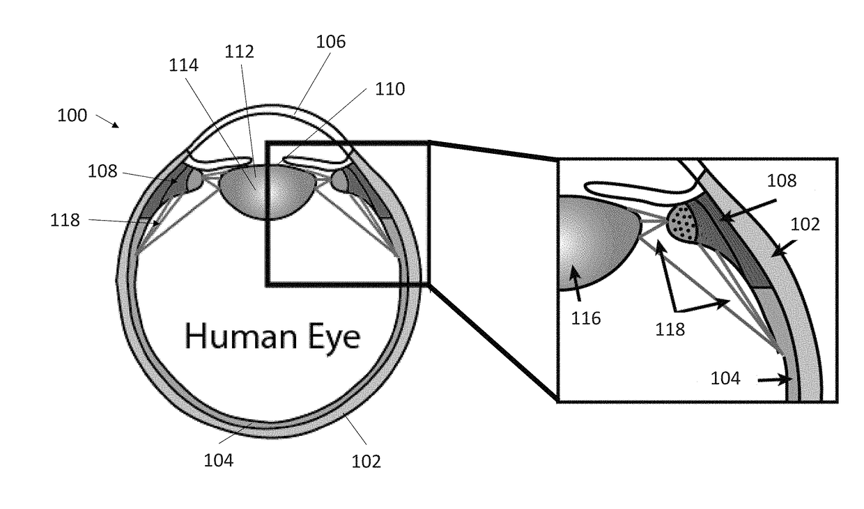
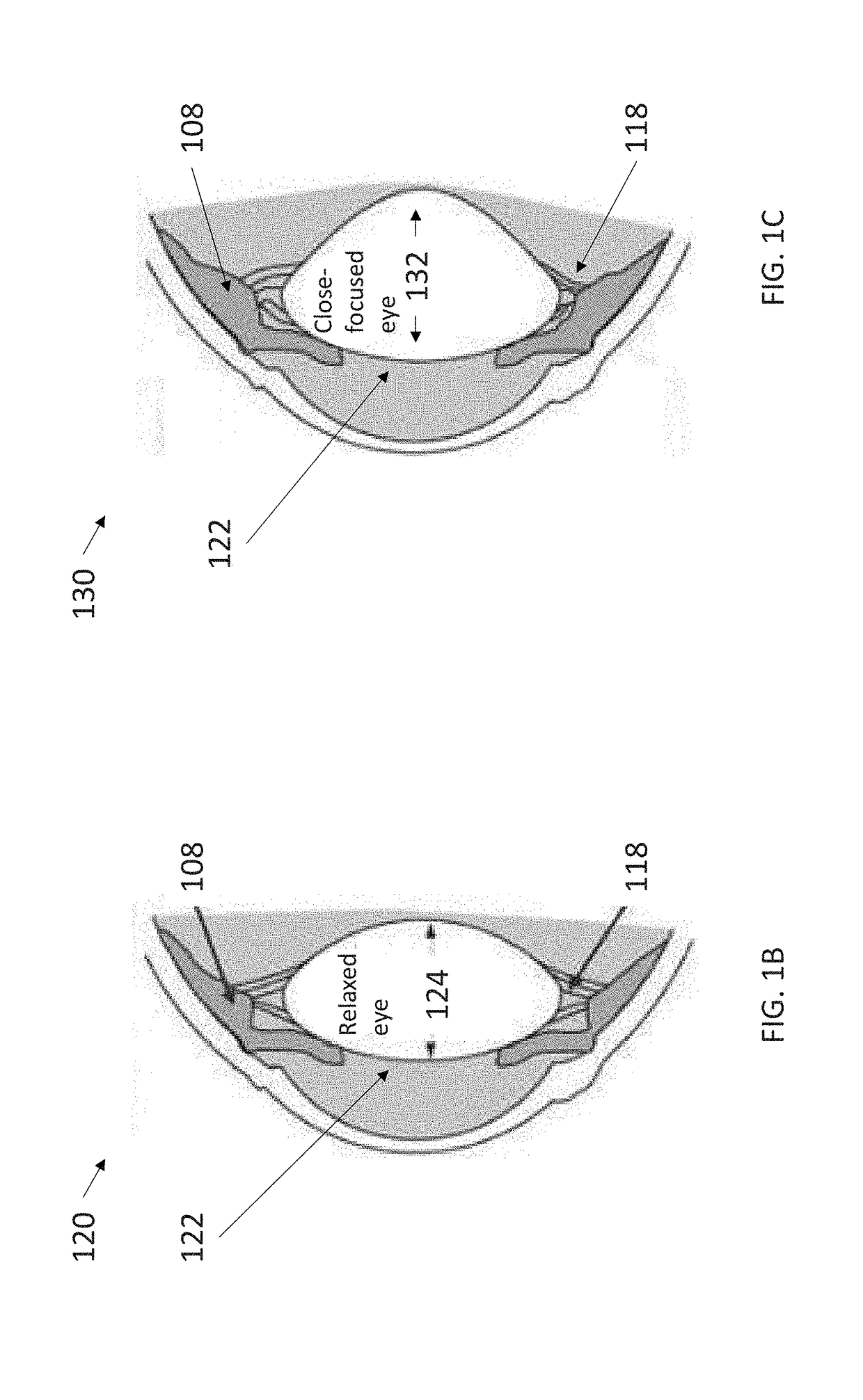
[0158]Accommodation of a human eye occurs through a change or deformation of the ocular lens when the eye transitions from distant focus to near focus. This lens change is caused by contraction of intraocular ciliary muscles that make up the ciliary body, which relieves tension on the lens through suspensory zonule fibers and allows the thickness and surface curvature of the lens to increase. The ciliary muscle can have a ring-shape and can be composed of three uniquely oriented ciliary fiber groups that contract toward the center and anterior of the eye. These t...


PUM


Abstract

Disclosed are systems, devices and methods for a modeling of ocular structures involved in ocular accommodation and use of a multi-component Finite Element Model (FEM).

Description

CROSS REFERENCE TO RELATED APPLICATIONS[0001]This application claims priority to U.S. Appl. No. 62 / 356,457, filed Jun. 29, 2016 and titled “3-DIMENSIONAL MODEL CREATION USING WHOLE EYE FINITE ELEMENT MODELING OF HUMAN OCULAR STRUCTURES FOR USE IN CREATING ACCURATE SIMULATION OF OCULAR BIOMECHANICS,” the entire contents and disclosure of which is hereby incorporated by reference.[0002]This application is related to the subject matter disclosed in U.S. Appl. No. 61 / 798,379, filed Mar. 15, 2013; U.S. Appl. No. 60 / 662,026, filed Mar. 15, 2005; U.S. application Ser. No. 11 / 376,969, filed Mar. 15, 2006; U.S. Appl. No. 60 / 842,270, filed Sep. 5, 2006; U.S. Appl. No. 60 / 865,314, filed Nov. 10, 2006; U.S. Appl. No. 60 / 857,821, filed Nov. 10, 2006; U.S. application Ser. No. 11 / 850,407, filed Sep. 5, 2007; U.S. application Ser. No. 11 / 938,489, filed Nov. 12, 2007; U.S. application Ser. No. 12 / 958,037, filed Dec. 1, 2010; U.S. application Ser. No. 13 / 342,441, filed Jan. 3, 2012; U.S. application...

Claims


Application Information

Patent Timeline
Patent Type & Authority Applications(United States)
IPC IPC(8): G06F19/00G06T17/20G06F17/50
CPCG06F19/3437G16H50/50G06T17/20G06F17/5018G06F30/23A61B3/00
Inventor HIPSLEY, ANNMARIEBLEMKER, SYLVIA S.KNAUS, KATIE R.
Owner ACE VISION GRP INC
Why Eureka
Try Eureka