Digital pathology system and associated workflow for providing visualized whole-slide image analysis

a pathology system and workflow technology, applied in image analysis, instruments, image enhancement, etc., can solve the problems of reducing the ability for real-time diagnostic and interaction execution, computationally intensive digital image analysis, and requiring significant storag

Active Publication Date: 2019-08-01
VENTANA MEDICAL SYST INC
View PDF0 Cites 4 Cited by
  • Summary
  • Abstract
  • Description
  • Claims
  • Application Information

AI Technical Summary

Benefits of technology

[0008]It is anticipated that whole-slide image analyses will become widely adapted in automated image analyses for therapeutic, prognostic, or diagnostic decision support. As a result, the present disclosure describes an efficient whole-slide image analysis workflow for use in a digital pathology system to perform quantitative analysis of the entire tissue slides as well as intuitive visualization.

Problems solved by technology

Current digital pathology image analysis systems are increasingly expected to analyze large amounts of high-resolution digital images of tissue slides or “virtual slides.” The digital image analysis has proven to be computationally intensive, requiring significant storage, and lessening the ability for real time diagnostic and interaction execution.
Despite their potential advantages, current digital pathology systems have not achieved the desired efficiency in providing an expeditious, real time quantitative analysis of entire tissue slides as well as intuitive visualization thereof.

Method used


Image

  • Digital pathology system and associated workflow for providing visualized whole-slide image analysis
  • Digital pathology system and associated workflow for providing visualized whole-slide image analysis
  • Digital pathology system and associated workflow for providing visualized whole-slide image analysis

Examples


Embodiment Construction

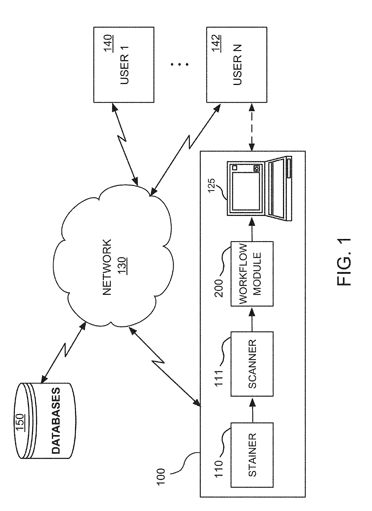
[0040]FIG. 1 illustrates a computer-based digital pathology system 100 that operates in a network environment for providing a visual quantitative analysis of a whole-slide image as well as intuitive visualization of the quantification of biomarker expressions, in accordance with one embodiment of the present disclosure. The digital pathology system 100 interfaces with a plurality of client computer systems (or user stations) 140, 142 over a network 130.

[0041]The digital pathology system 100 may include, among other things, a stainer 110, a scanner 111, a workflow module 200 and a processor or computer 125. The users of the client computer systems 140, 142, such as pathologists, histotechnologists, or like professionals, may be able to access, view, and interface with the outputs of the scanner 111 and workflow module 200 on a real time basis, either remotely or locally. These outputs may alternatively be stored and accessed on networked databases 150.

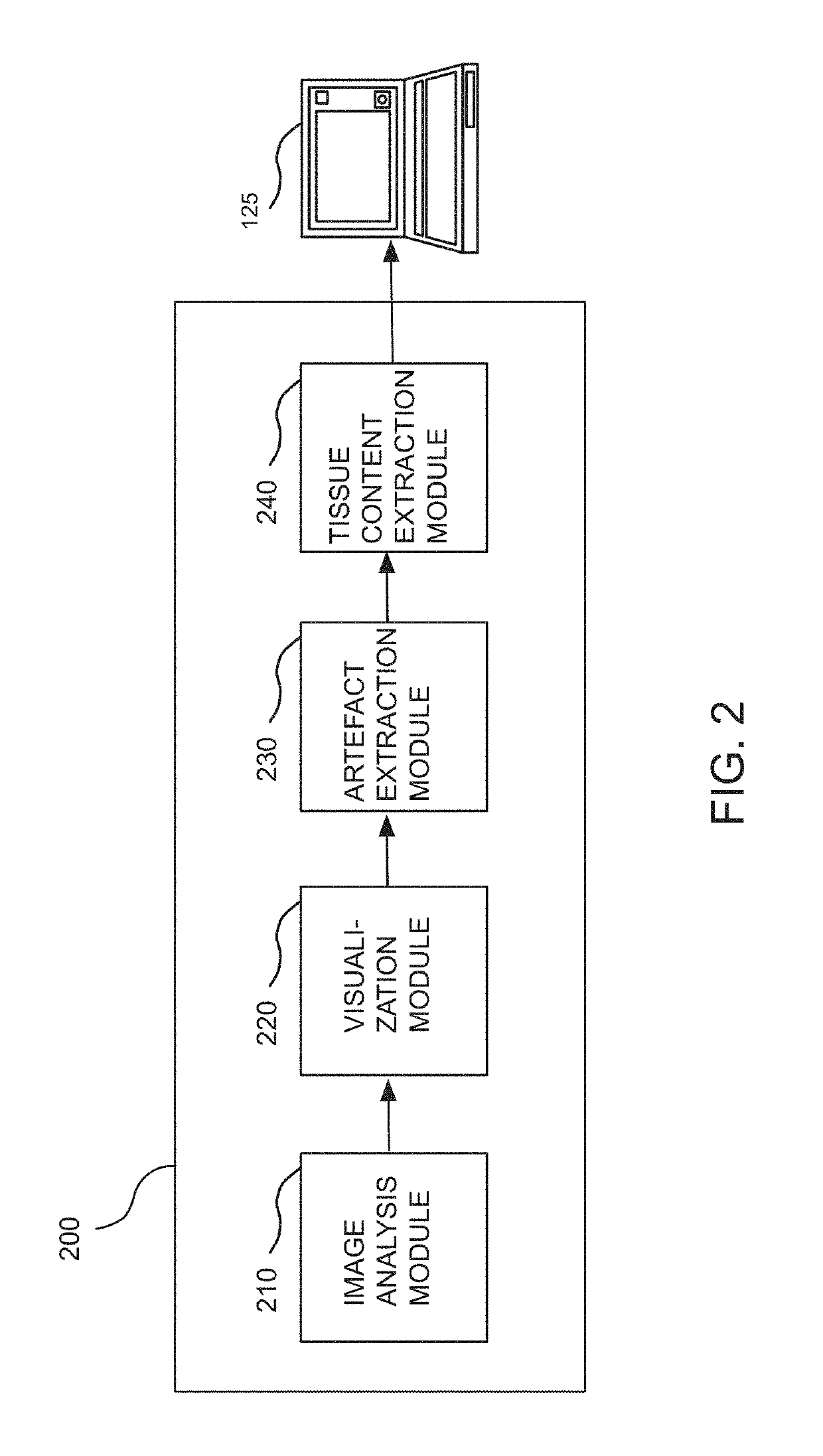
[0042]As further detailed in FIG...


PUM


Abstract

A digital pathology system and associated method and computer program product provide a quantitative analysis of entire tissue slides as well as intuitive, effective, fast, and precise quantification of biomarker expressions across relevant areas of the entire tissue slides. The digital pathology system enables a novel workflow that allows the user to efficiently outline clinically relevant morphology in its entirety, including solid tumor areas. Quantitative analysis results are then efficiently and intuitively provided to the user for all tissue content (i.e., millions of cells) within seconds. This efficiency is made possible by a pre-computation step that computes and stores image analysis results for later retrieval. Visualizing vast amount of data effectively is another component of the system that provides information and confidence to the user about the biomarker expression levels.

Description

RELATED APPLICATIONS[0001]This patent application is a continuation of International Patent Application No. PCT / EP2017 / 075113 filed Oct. 4, 2017, which claims priority to and the benefit of U.S. Provisional Application No. 62 / 405,808, filed Oct. 7, 2016, and entitled “Digital Pathology System and Associated Workflow for Providing Visualized Whole-Slide Image Analysis.” The contents of all the aforementioned applications are hereby incorporated by reference in their entirety into this disclosure.TECHNICAL FIELD[0002]The present disclosure generally relates to a digital pathology system that hosts a workflow for providing a quantitative analysis of whole-slide images as well as intuitive visualization thereof.BACKGROUND[0003]Digital pathology equipment is often used to produce digital images of microscope slides. Pathologists and histotechnologists often visually examine the digital images to obtain information about tissue samples and to identify the most appropriate treatment to imp...

Claims


Application Information

Patent Timeline
Patent Type & Authority Applications(United States)
IPC IPC(8): G06T7/00G06K9/00
CPCG06T7/0012G06K9/00127G06T2207/30096G06T2207/30168G06T2207/10056G06T2207/30024G06T7/11G06T2207/30204G06V20/69
Inventor BARNES, MICHAELBREDNO, JOERGCHUKKA, SRINIVASGUETTER, CHRISTOPHLORSAKUL, AURANUCHSARKAR, ANINDYASUZUE, ELLEN
Owner VENTANA MEDICAL SYST INC
Why Eureka
Try Eureka