Instrument for repairing an atrioventricular heart valve

a heart valve and atrioventricular technology, applied in the field of minimally invasive surgical and interventional cardiology devices for heart valve repair, can solve the problems of long time damage, high dependence on the success of the operation, and serious heart failure, and achieve reliable and well tissue-compliant repair, easy implantation, and the effect of good tissue-compatibility

Pending Publication Date: 2022-06-16
COREMEDIC GMBH
View PDF0 Cites 0 Cited by
  • Summary
  • Abstract
  • Description
  • Claims
  • Application Information

AI Technical Summary

Benefits of technology

[0008]It is an object of the present invention to provide an instrument for repairing an atrioventricular heart valve, in particular the mitral heart valve or also the tricuspid heart valve, and an according method, which instrument and method overcome drawbacks of prior art devices and methods and which ensure easy implantation, are suited also for transcatheter procedures and provide a reliable and well tissue-compliant repair. The transcatheter procedure is carried out in a minimally invasive manner on the beating heart, the leaflet has to be held still for the operation, and the instrument and method should provide solutions for this. Especially, it should easily be possible to check the orientation and placement of the instrument using echocardiopathy.

Problems solved by technology

Prolapses of a leaflet of the mitral valve into the left atrium and resulting valve insufficiency can cause serious dysfunctions of the heart.
A first disadvantage of such a repair process is that it is only possible while the heart is inactive, thus the surgical repair demands that the heart is stopped and drained of blood, while a cardiopulmonary bypass is used.
A second disadvantage is that the success of the operation depends strongly on the skill of the surgeon.
A further disadvantage is that the fibers sutured to the leaflet may cause long-time damage.
Most laparoscopic and minimally invasive techniques and tools that have been developed are not usable for the requirements of mitral valve repair on a beating heart.
The complete mechanism is time consuming and prone to error.
In addition, methods that are based on sutures feature the disadvantage that suturing in minimally invasive setups is difficult, and therefore sufficiently reliable stability is often difficult to achieve.
It has been found that this structure causes problems during orientation and placement of the instrument using echocardiography.
Furthermore, the control mechanism is rather complicated.

Method used


Image

  • Instrument for repairing an atrioventricular heart valve
  • Instrument for repairing an atrioventricular heart valve
  • Instrument for repairing an atrioventricular heart valve

Examples


Embodiment Construction

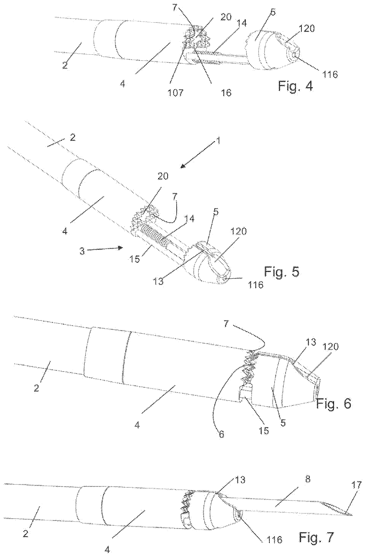
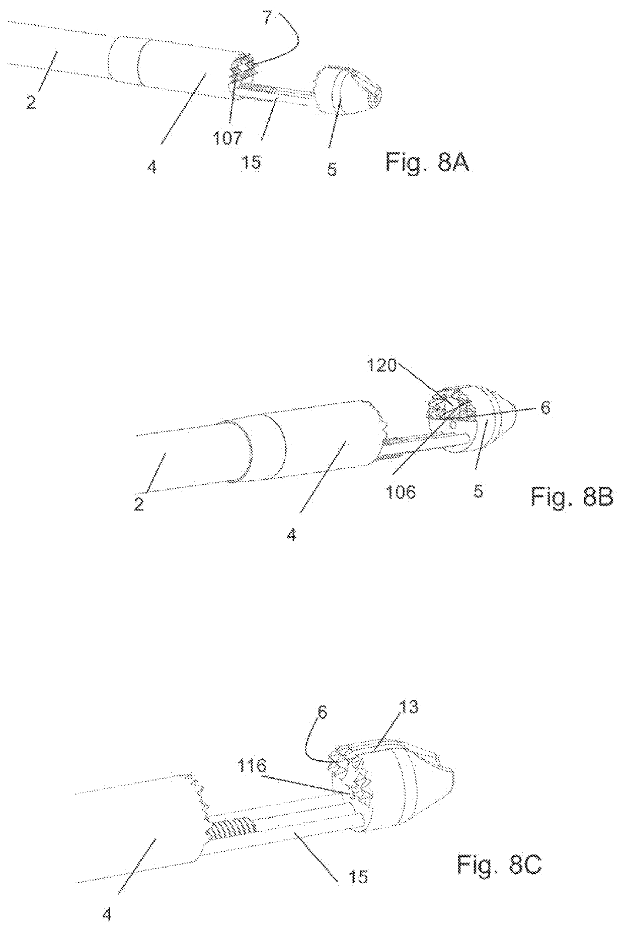
[0104]The following more detailed description of the embodiments of the instrument is a representative of exemplary embodiments of the technology, wherein similar parts are designated by same numerals throughout. Standard medical planes of reference and descriptive terminology are employed in this specification. In particular, proximal means toward the trunk, or, in the case of an inanimate object, toward a user and distal means away from the trunk, or, in the case of an inanimate object, away from a user.

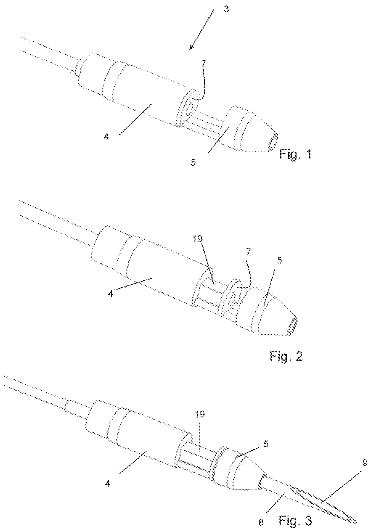
[0105]FIGS. 1 to 3 show one exemplary embodiment of a grabbing structure being part of an instrument for repairing an atrioventricular heart valve. FIG. 1 shows a grabbing structure 3 including a main body 4 and a jaw 5 in an open position. The grabbing structure may be formed as a bullet tip grasper (bullet tip grabber). The grabbing structure may have a length of 0.75 to 1.2 cm. The jaw may be formed as a distal nosecone tip able to be advanced over two guiding rails to create a ...


PUM


Abstract

An instrument for repairing an atrioventricular heart valve, mitral heart valve or tricuspid heart valve includes a first tube having a tube axis defining an axial direction. A leaflet grabbing structure is arranged at a distal part of the first tube and contiguous to the first tube. The leaflet grabbing structure has a main body and a jaw, the jaw being movable relative to the main body. The jaw has a first proximally facing abutment surface and the main body has a second, distally facing abutment surface. The instrument has a needle that is cannulated and forms an inner tube. The needle is releasable from the first tube and movable relative to the main body in axial directions. The leaflet grabbing structure clamps a leaflet of the heart valve between the first and second abutment surfaces, while the jaw allows the needle to extend therethrough.

Description

BACKGROUND OF THE INVENTIONField of the Invention[0001]The invention is in the field of minimally invasive surgical and interventional cardiology devices for heart valve repair. It particularly relates to an instrument for repairing an atrioventricular heart valve, mitral heart valve or also the tricuspid heart valve in a minimally invasive manner, and to an according method.Description of Related Art[0002]Prolapses of a leaflet of the mitral valve into the left atrium and resulting valve insufficiency can cause serious dysfunctions of the heart. One reason for such prolapse are damaged tendons (chordae tendineae) that connect the leaflets of the mitral valve to the papillary muscle through the left ventricle. Such damage may, for example, be a result of a myocardial infarction, tissue degeneration or infectious disease.[0003]A repair of such a prolapse demands the leaflet or leaflets to be re-connected to the papillary muscle, for example by synthetic fibers, such as Gore-Tex® fibe...

Claims


Application Information

Patent Timeline
Patent Type & Authority Applications(United States)
IPC IPC(8): A61F2/24
CPCA61F2/2466A61F2/2457A61B2017/00292A61B2017/003A61B2017/06052A61B17/06A61B2017/00738A61B2017/0409A61B2017/00783
Inventor BAUER, THOMASSIRONI, TOMMASOROTH, MICHAELSCHAFFNER, SILVIOGUIDOTTI, ANDREAZARBATANY, DAVIDFLORES, JESUSSTEFOPOULOS, GEORGIOSVICENTINI, LUCAARDUINI, MATTIA
Owner COREMEDIC GMBH
Why Eureka
Try Eureka