Compressible tissue anchor assemblies

a tissue anchor and compression technology, applied in the field of tissue anchors, can solve problems such as over-compression of underlying tissue, and achieve the effect of constant force against tissu

Active Publication Date: 2010-06-15
USGI MEDICAL
View PDF328 Cites 230 Cited by
  • Summary
  • Abstract
  • Description
  • Claims
  • Application Information

AI Technical Summary

Benefits of technology

[0011]In securing the tissue folds or anchoring to or from these tissue folds or plications, over-compression of the tissue directly underlying the tissue anchors is preferably avoided. Over-compression of the underlying tissue may occur if the anchor compresses the tissue to such a degree that tissue necrosis or cutting of the underlying muscularis or serosal tissue by the anchor occurs. Accordingly, a tissue anchor is preferably configured to maintain or secure a tissue plication yet still allow for adequate blood flow to occur within the tissue underlying the anchor. As such, the tissue anchor is preferably configured to accommodate a range of deflections due to various movements of the tissue due to, e.g., peristalsis, patient movement, weight of the gastrointestinal organ itself, etc., while maintaining or exerting a substantially constant force against the tissue.
[0012]A particular type of anchor which may be utilized is a reconfigurable “basket”-type anchor generally having a number of configurable struts or legs extending between at least two collars or bushing members. This anchor may have a low-profile delivery configuration and a radially expanded anchoring configuration. When expanded, each arm of the anchor may be separated from one another by a spacing or opening. The spacing is preferably created to minimize the contact area between the anchor body and the underlying tissue surface to allow for greater blood flow in the tissue and to inhibit necrosis of the tissue.
[0016]One method for limiting the loading or pre-load force upon an anchor may involve including a post or stop in the anchor body which limits the proximal deflection of the distal collar and thus prevents over-compression of the anchor against the tissue. Another variation may utilize friction-producing regions within the anchor delivery catheter. As the anchor is tensioned, various regions may produce frictional forces which vary in accordance to the degree of anchor deflection. A change in the detected frictional force may thus be utilized to indicate that anchor has been configured within an optimal range of deflections.
[0018]Yet another variation may include configuring the proximal collar of the anchor to prevent the passage of stop member contained within the anchor. thus, the length of suture extending from the stop member to the attachment point within the anchor may be of a predetermined length such that when the stop member is seated against the proximal collar, the suture length may compress the anchor into a predetermined deflection level. This deflection level may be preset to configure the anchor to any desired configuration, as described above.

Problems solved by technology

Over-compression of the underlying tissue may occur if the anchor compresses the tissue to such a degree that tissue necrosis or cutting of the underlying muscularis or serosal tissue by the anchor occurs.

Method used


Image

  • Compressible tissue anchor assemblies
  • Compressible tissue anchor assemblies
  • Compressible tissue anchor assemblies

Examples


Embodiment Construction

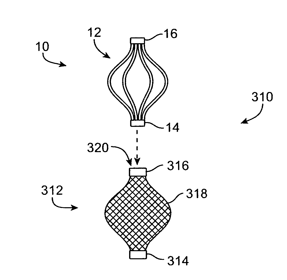
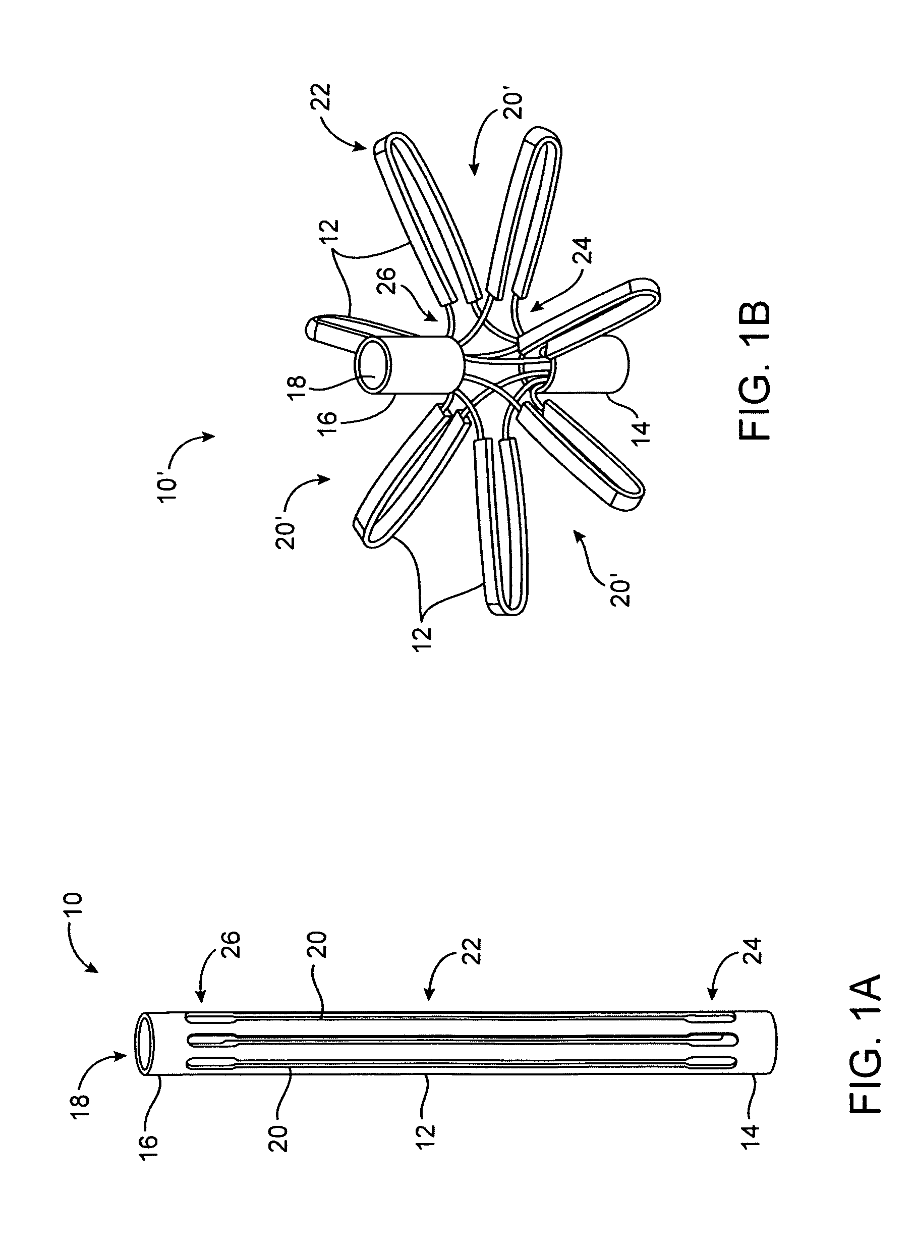
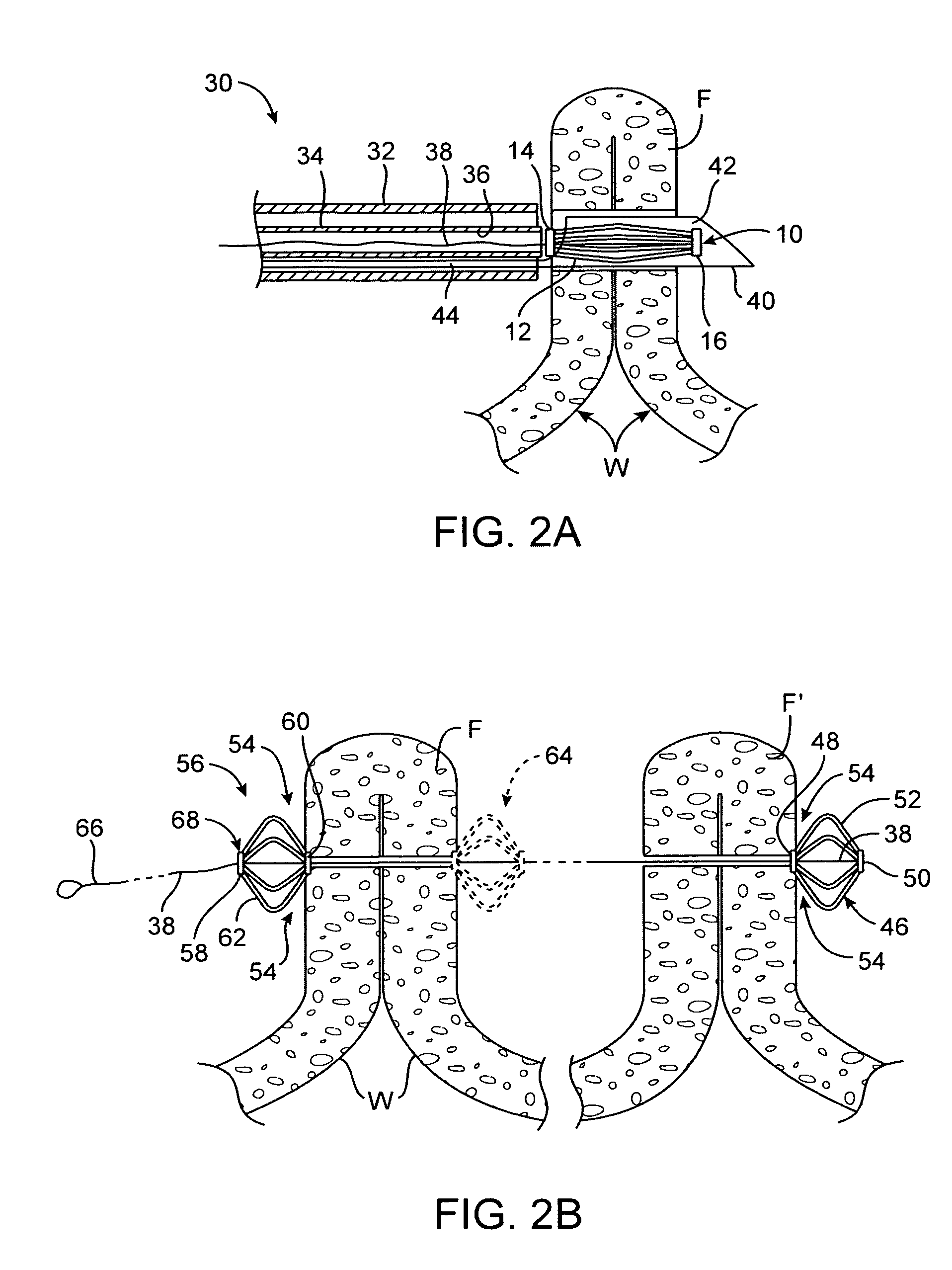
[0060]Generally, in creating and securing a plication within a body lumen of a patient, various methods and devices may be implemented. The anchoring and securement devices may be delivered and positioned via an endoscopic apparatus that engages a tissue wall of the gastrointestinal lumen, creates one or more tissue folds, and disposes one or more of the anchors through the tissue fold(s).

[0061]In securing the tissue folds or anchoring to or from these tissue folds or plications, over-compression of the tissue directly underlying the tissue anchors is preferably avoided. Over-compression of the underlying tissue may occur if the anchor compresses the tissue to such a degree that tissue necrosis or cutting of the underlying muscularis or serosal tissue by the anchor occurs. The anchor preferably exerts a force, e.g., about 0.1-0.5 lbs, sufficient to maintain or secure a tissue plication yet still allows for adequate blood flow to occur within the tissue underlying the anchor. Accordi...


PUM


Abstract

Apparatus & methods for optimizing anchoring force are described herein. In securing tissue folds, over-compression of the tissue directly underlying the anchors is avoided by utilizing tissue anchors having expandable arms configured to minimize contact area between the anchor and tissue. When the anchor is in its expanded configuration, a load is applied to the anchor until it is optimally configured to accommodate a range of deflections while the anchor itself exerts a substantially constant force against the tissue. Various devices, e.g., stops, spring members, fuses, strain gauges, etc., can be used to indicate when the anchor has been deflected to a predetermined level within the optimal range. Moreover, other factors to affect the anchor characteristics include, e.g., varying the number of arms or struts of the anchor, positioning of the arms, configuration of the arms, the length of the collars, etc.

Description

CROSS-REFERENCES TO RELATED APPLICATIONS[0001]This application is a continuation-in-part of U.S. patent application Ser. No. 10 / 865,243, filed Jun. 9, 2004, which is incorporated herein by reference in its entirety.BACKGROUND OF THE INVENTIONField of the Invention[0002]The present invention relates to improved tissue anchors for securement against tissue. More particularly, the present invention relates to tissue anchors which are deployable into or against tissue for securing portions thereof.[0003]Morbid obesity is a serious medical condition pervasive in the United States and other countries. Its complications include hypertension, diabetes, coronary artery disease, stroke, congestive heart failure, multiple orthopedic problems and pulmonary insufficiency with markedly decreased life expectancy.[0004]A number of surgical techniques have been developed to treat morbid obesity, e.g., bypassing an absorptive surface of the small intestine, or reducing the stomach size. However, many...

Claims


Application Information

Patent Timeline
Patent Type & Authority Patents(United States)
IPC IPC(8): A61B17/04A61B17/00A61B17/06A61B17/064A61B17/08A61B19/00
CPCA61B17/0401A61B17/0482A61B17/0644A61B17/0469A61B2019/307A61B2017/0454A61B2017/0464A61B2017/0496A61B2017/06052A61B2017/00867A61B2090/037
Inventor EWERS, RICHARD C.VONG, SHIRLEYSAADAT, VAHID
Owner USGI MEDICAL
Why Eureka
Try Eureka