Anthropomorphic phantom for medical imaging systems

a technology of medical imaging and phantoms, applied in the field of medical imaging techniques, can solve the problems of not having space available in all the other organs, a prior-art anthropomorphic torso phantom is not useful in medical imaging researches for tumors and lesions, and a prior-art thorax phantom is not useful in tumors and lesions

Inactive Publication Date: 2015-03-03
INST NUCLEAR ENERGY RES ROCAEC
View PDF4 Cites 43 Cited by
  • Summary
  • Abstract
  • Description
  • Claims
  • Application Information

AI Technical Summary

Benefits of technology

[0008]The object of the present invention is to provide an anthropomorphic phantom for medical imaging systems, that is configured with organ models that are designed to mimic the three-dimensional shape of human organs while allowing solutions of contrast agents or medicines to be filled therein, and also by the arrangement of a plate-like first connecting element inside each of the corresponding organ models, lesion phantoms that are used for simulating tumors are fixedly secured inside the corresponding organ model by the use of a plurality of first connection holes that are arranged distributing on the first connecting element. Moreover, by the distribution of those first connection holes, not only each of the lesion phantoms can be arranged at any position inside the corresponding organ model at will as required, but also there can be more than one lesion phantoms either of the same size or of different sizes to be arranged inside the same organ model simultaneously. Thereby, the anthropomorphic phantom can very adaptive for simulating real lesions in human body.
[0009]Another object of the invention to provide an anthropomorphic phantom with modularized design, by that an organ model can first be fixedly secured to a second connecting element of a module structure while the whole module structure with the organ model can be assembled into a torso-like humanoid housing of the anthropomorphic phantom by the use of a handle element. Thereby, not only the assembling of organ model to the torso-like humanoid housing can be simplified, but also any experimental error causing by contamination of contrast agents or medicines can be prevented.

Problems solved by technology

Moreover, it is disadvantage in that except for a specific position on the breast, there is no space available in all the other organs that can be used for allowing any spherical tumor / lesion phantom to be arranged thereat.
Thus, the aforesaid prior-art anthropomorphic torso phantom is not useful in medical imaging researches for tumors and lesions.
Thus, the aforesaid prior-art thorax phantom is also not useful in medical imaging researches for tumors and lesions.
In addition to the sizes as well as the positions of those cylinders are fixed and can not be changed, the appearance of the cylinder too simple to mimic a real lesion.
Although the aforesaid prior-art multicontrast / resolution phantom can be used for generating lesion images of different contrasts and resolutions, it is still not a complete 3D liver and also is disadvantageous in that the sizes as well as the positions of those acrylic lesion phantoms are fixed and can not be changed.

Method used


Image

  • Anthropomorphic phantom for medical imaging systems
  • Anthropomorphic phantom for medical imaging systems
  • Anthropomorphic phantom for medical imaging systems

Examples


Embodiment Construction

[0029]For your esteemed members of reviewing committee to further understand and recognize the fulfilled functions and structural characteristics of the invention, several exemplary embodiments cooperating with detailed description are presented as the follows.

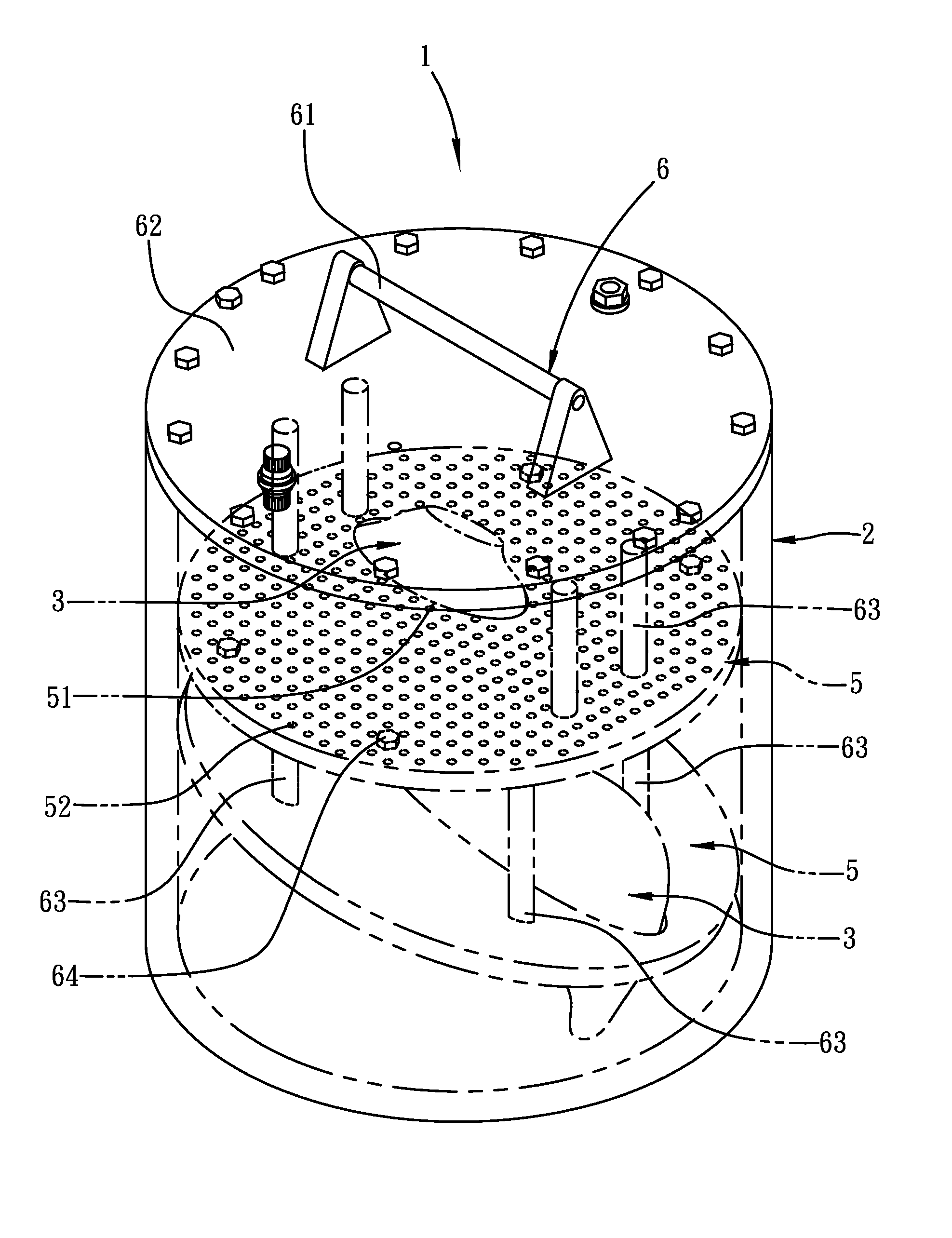
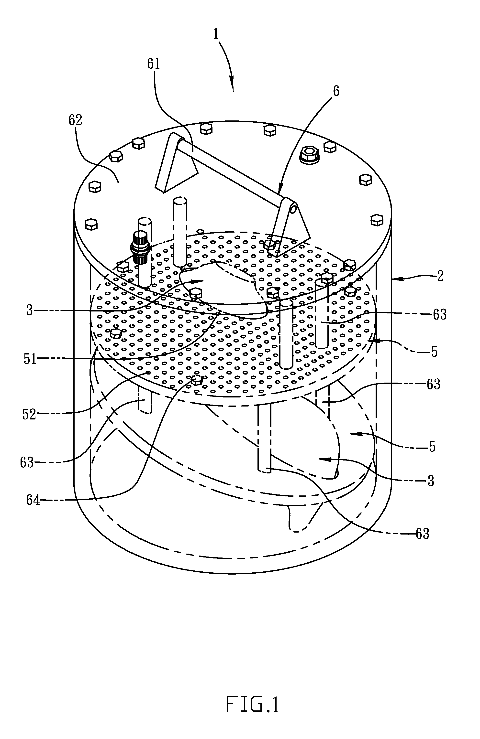
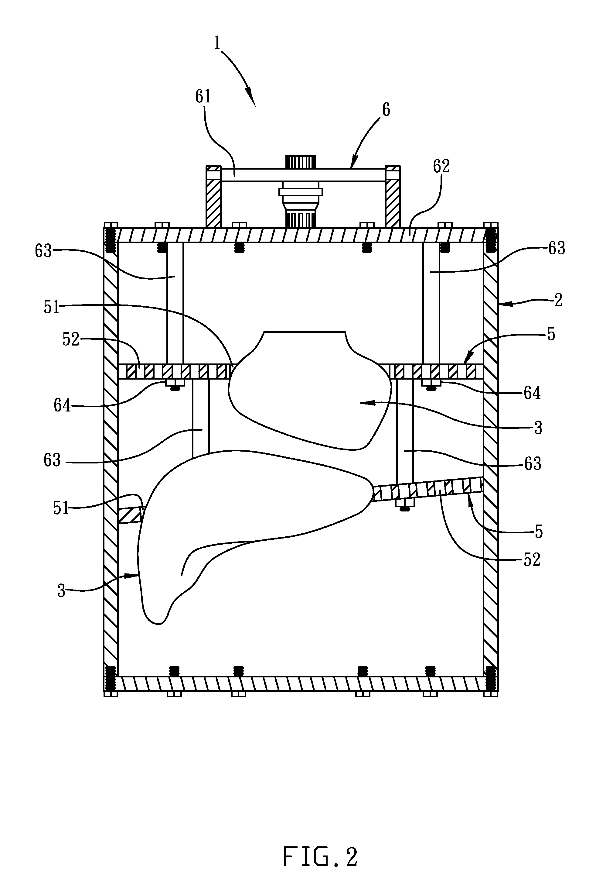
[0030]FIG. 1 is a perspective diagram showing an anthropomorphic phantom with cylinder-shape housing according to an embodiment of the present invention. FIG. 2 is a cross sectional view of FIG. 1. FIG. 3 is a three-dimensional diagram showing the structure inside the humanoid housing of an anthropomorphic phantom according to an embodiment of the present invention. FIG. 4 is a schematic diagram showing a liver-shape organ model that is adapted for an anthropomorphic phantom of the present invention. FIG. 5 is a perspective view of a liver-shape organ model that is adapted for an anthropomorphic phantom of the present invention.

[0031]The anthropomorphic phantom 1 of the present invention comprises: a humanoid housing 2, at lea...


PUM

PropertyMeasurementUnit
diameteraaaaaaaaaa
structureaaaaaaaaaa
sizesaaaaaaaaaa

Abstract

An anthropomorphic phantom for medical imaging systems is disclosed, which is configured with organ models designed to mimic the three-dimensional shape of human organs, and also by the arrangement of plate-like first connecting elements inside their corresponding organ models, lesion phantoms for simulating tumors can be fixedly secured inside the corresponding organ model using a plurality of first connection holes formed on the first connecting element. Therefore, by the distribution of those first connection holes, not only the lesion phantoms can be arranged at any position inside the corresponding organ model at will as required, but also by modularized design of the anthropomorphic phantom, organ models can first be assembled to form a module structure while the whole module structure with the organ model can be moved into and assembled easily with a torso-like humanoid housing of the anthropomorphic phantom by the use of a handle element.

Description

FIELD OF THE INVENTION[0001]The present invention relates to a medical imaging technique, and more particularly, to an anthropomorphic phantom for medical imaging systems.BACKGROUND OF THE INVENTION[0002]Notably, there are already many anthropomorphic phantoms available on the market. One of which is an anthropomorphic torso phantom by Data Spectrum Corporation, as shown in FIG. 6. In FIG. 6, although the anthropomorphic torso phantom is configured with a heart, a breast, a portion of lung, a liver and a spinal cord, only the heart, breast, liver and torso can be filled by a solution of contrast agent or medicine. Moreover, it is disadvantage in that except for a specific position on the breast, there is no space available in all the other organs that can be used for allowing any spherical tumor / lesion phantom to be arranged thereat. Thus, the aforesaid prior-art anthropomorphic torso phantom is not useful in medical imaging researches for tumors and lesions.[0003]Another such anthr...

Claims


Application Information

Patent Timeline
Patent Type & Authority Patents(United States)
IPC IPC(8): G01N29/30G09B23/28G09B23/30
CPCG09B23/30G09B23/286A61B6/583
Inventor NI, YU-CHINGHSU, SHIANG-LINJAN, MEEI-LING
Owner INST NUCLEAR ENERGY RES ROCAEC
Why Eureka
Try Eureka