
Dose guided deformable image registration
An image registration and deformation map technology, applied in the field of radiotherapy, can solve the problems of dose error influence, DIR geometric error, etc.
Patent Information
- Authority / Receiving Office
- CN · China
- Patent Type
- Applications(China)
- Current Assignee / Owner
- Publication Date
- 2022-04-12
Smart Images
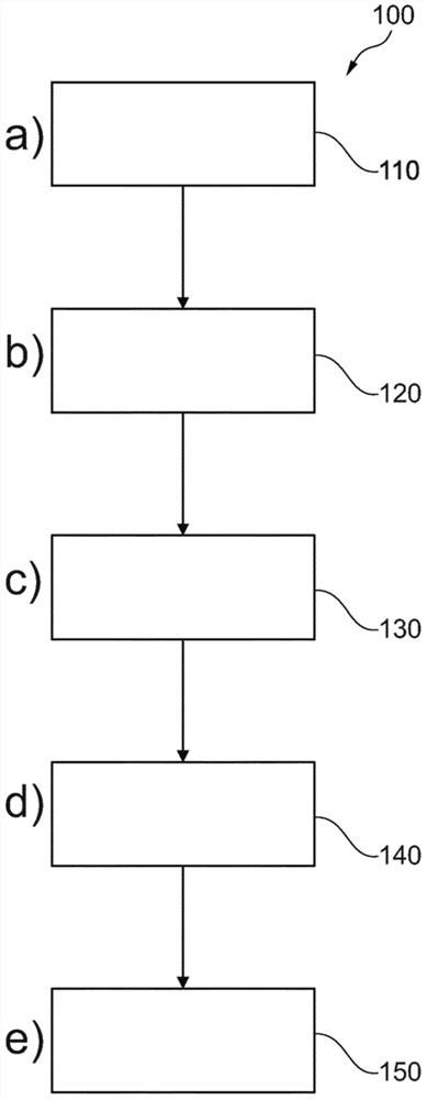
Figure 1 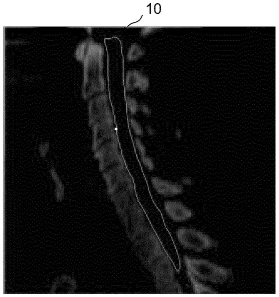
Figure 2 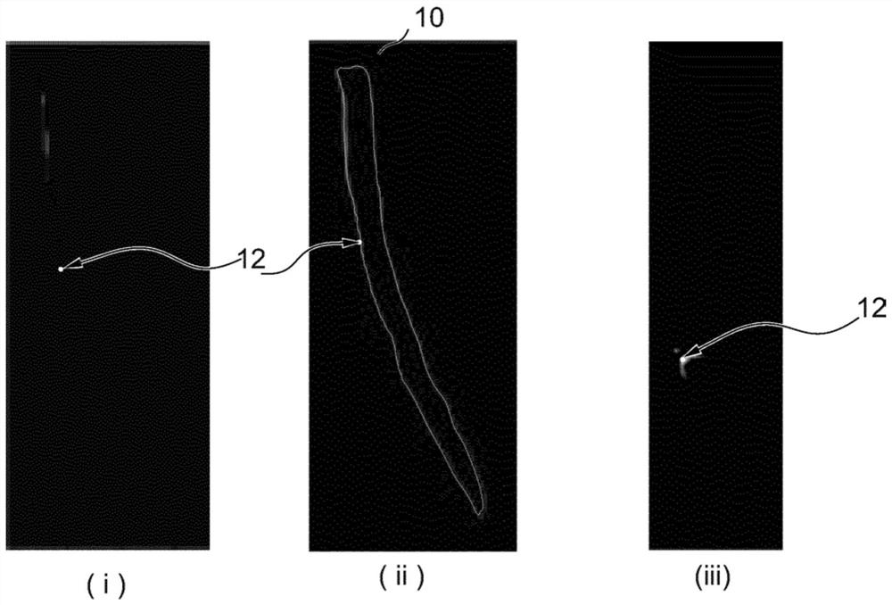
Figure 3
Abstract
Description
technical field
[0001] The present invention relates to radiation therapy. In particular, the present invention relates to a radiotherapy planning method and a radiotherapy planning device. The invention also relates to a computer program element. Background technique
[0002] Deformable image registration (DIR) is the key to radiotherapy, adaptive planning, and composite planning, and the trend of large segmentation intensifies the requirement for accuracy.
[0003] In radiation therapy, DIR is used to estimate the deformation between images used for treatment planning and / or guidance, and then to map the outline of a region of interest (ROI) for radiation therapy planning, mapping computed tomography (CT) density to another A modality such as cone beam CT (CBCT) in adaptive RT, magnetic resonance (MR) in MR-only simulation (MRCAT) to create a virtual CT or attenuation map, and to accumulate dose. Radiation therapy planning ROI is subject to dose objective constraints in...
Examples
Embodiment Construction
[0051] figure 1 A radiation therapy planning method 100 for registering at least two three-dimensional medical images of a patient is shown. The method includes the following steps:
[0052] In step 110 , step a), a dose profile to be delivered during one or more treatment sessions according to the radiotherapy plan is received.
[0053] In step 120, ie step b), a contour delineating at least one region of interest ROI of at least one of the medical images is received. Automatic segmentation tools can be used to delineate at least one ROI. Alternatively or additionally, a manual segmentation tool may be used which allows the user to manually adjust image features, in particular segmentation contours and / or landmark positions, when the medical image is displayed on the display. As an example, Figure 2A An example depicting a contour 10 of a patient's spinal cord, ie a ROI, is shown.
[0054] In step 130, step c), one or more treatment targets associated with at least one...