
Use of adipose tissue-derived stromal cells in spinal fusion
Patent Information
- Authority / Receiving Office
- US · United States
- Current Assignee / Owner
- Publication Date
- 2007-02-01
- Estimated Expiration
- Not applicable · inactive patent
Smart Images
Figure 1 
Figure 2 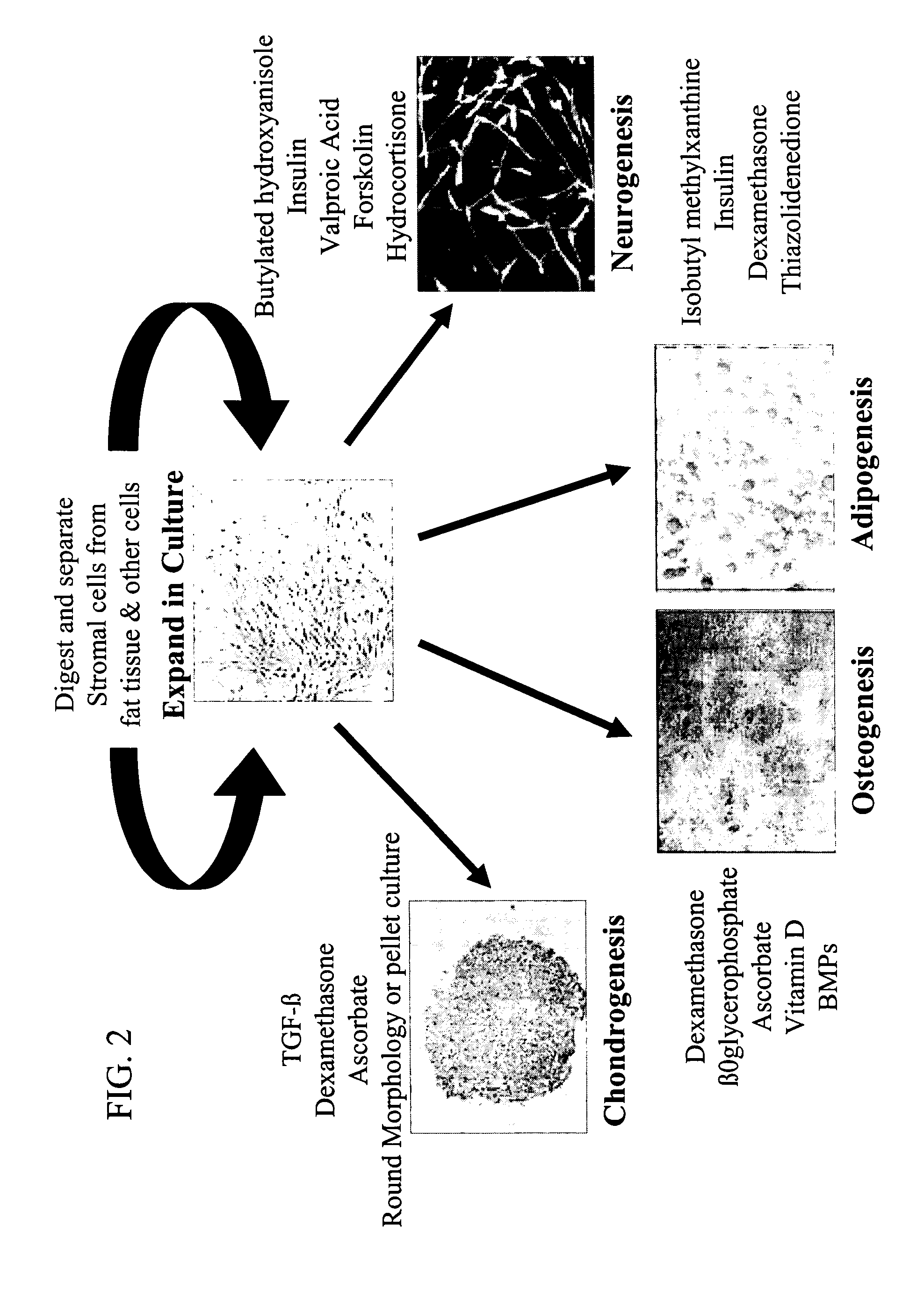
Figure 3
Abstract
Description
BACKGROUND OF THE INVENTION
[0001] There generally are two types of bone conditions: 1) non-metabolic bone conditions, such as bone fractures, bone / spinal deformation, osteosarcoma, myeloma, bone dysplasia and scoliosis, and 2) metabolic bone conditions, such as osteoporosis, osteomalacia, rickets, fibrous osteitis, renal bone dystrophy and Paget's disease of bone. Osteoporosis, a metabolic bone condition, is a systemic disease characterized by increased bone fragility and fracturability due to decreased bone mass and change in fine bone tissue structure, its major clinical symptoms including spinal kyphosis, and fractures of dorsolumbar bones, vertebral centra, femoral necks, lower end of radius, ribs, upper end of humerus, and others. In bone tissue, bone formation and destruction due to bone resorption occur constantly. Upon deterioration of the balance between bone formation and bone destruction due to bone resorption, a quantitative reduction in bone occurs. Traditionally, bone...
Examples
example 1
Alternatives to Autograft Bone in Spinal Fusion Surgery
[0160] Over 75% of the American population suffers from back pain. In some instances, underlying medical illnesses can contribute to back pain. These include scoliosis, spinal stenosis, degenerative disc disease, infectious processes, tumors, and trauma. For 1% of the population, the back pain is so severe that they are forced to go onto lifetime disability; an additional 1% of the population is incapacitated by back pain for a limited time period. The majority of the mammals with back pain are treated with conservative therapies; however, when modalities such as bed rest and medication fail, physicians often recommend spinal fusion surgery. The goal of this operation is to form ectopic bone between two or more adjacent vertebra, “fusing” them together into a solid structure. Immobilization of the vertebral joint reduces the pressure on nerve roots leaving the spinal cord and the resulting painful sensation. Surgeons use a post...
example 2
ADAS Cell Osteogenesis In Vitro
[0168] It has been demonstrated that human ADAS cells display an osteogenic phenotype in vitro when cultured in the presence of ascorbate, β-glycerophosphate, dexamethasone, and 1,25 dihydroxyvitamin D3 (Halvorsen et al., 2001, Tissue Eng. 7:729-41). Under these conditions, the ADAS cells mineralize their extracellular matrix as demonstrated by positive staining with either Alizarin Red or von Kossa for calcium phosphate deposition (FIG. 3).
[0169] It was observed that human ADAS cell osteogenesis over a 10-day period was accompanied by an increase in alkaline phosphatase activity. At the end of the culture period, osteogenic cells (mineralized) displayed a 3-fold higher level of alkaline phosphatase relative to cells maintained under control conditions (FIG. 4). Likewise, there was a time dependent increase in secreted levels of osteocalcin protein under osteogenic conditions. The ADAS cells expressed a number of gene markers consistent with an osteo...
example 3
ADAS Cells are Osteogenic In Vivo
[0171] To extend the in vitro findings, human ADAS cells were transplanted into immunodeficient SCID mice. The ADAS cells were loaded onto three cm3 cubes of hydroxyapatite / tricalcium phosphate (HA / TCP) scaffold and implanted subcutaneously. After a 6-week period, the implants were harvested, fixed, decalcified, and stained with Hematoxylin / Eosin or with human nuclear antigen specific antibodies (FIG. 5). Based on H&E staining, it was observed that new bone formed adjacent to the hydroxyapatite / tricalcium phosphate scaffold in the presence of the human ADAS cells. The human cells were identified within the bone based on immunofluorescent analysis with the human antigen specific antibody. In the presence of the scaffold alone (no cells), new bone did not form and no human cells were detected. These studies demonstrate that ADAS cells are capable of osteogenesis in vivo.