

Method for testing or screening protein synthesis inhibitors
Patent Information
- Authority / Receiving Office
- US · United States
- Current Assignee / Owner
- Publication Date
- 2010-10-28
Smart Images
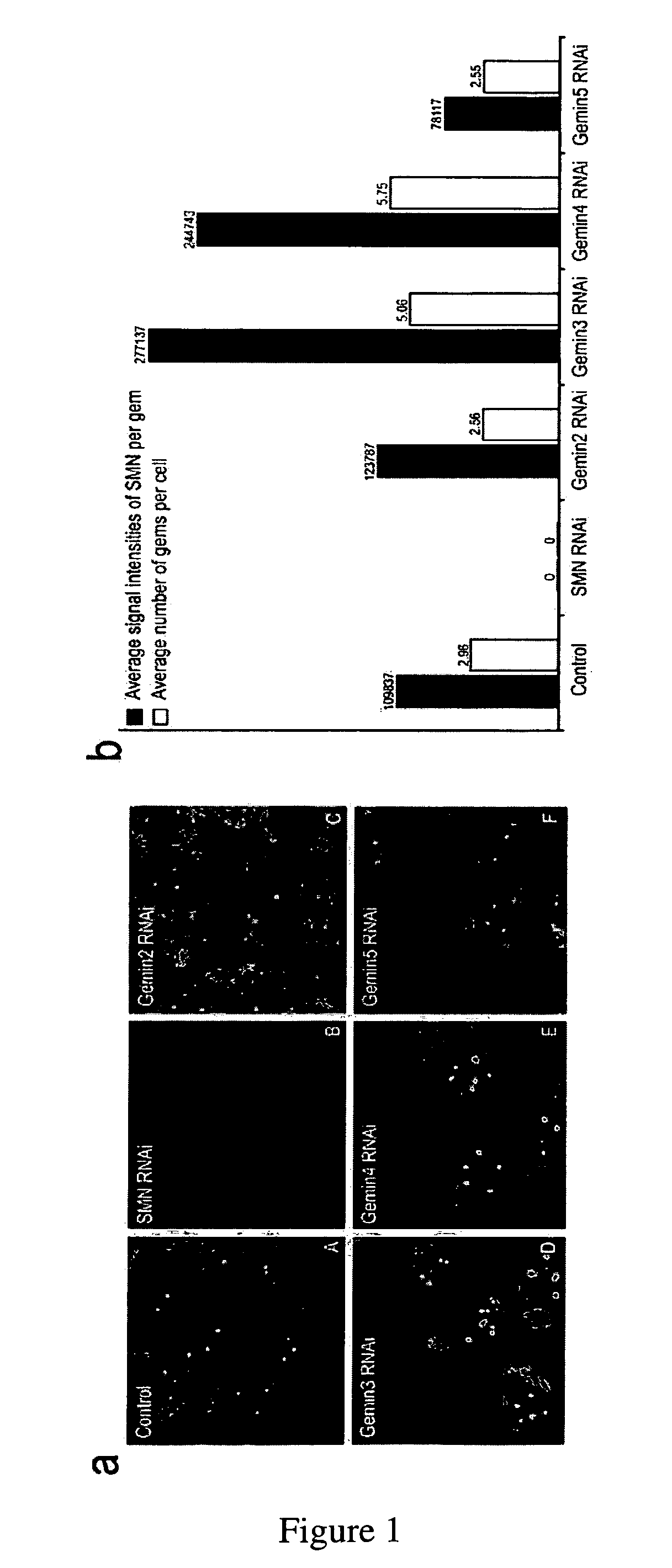
Figure 1 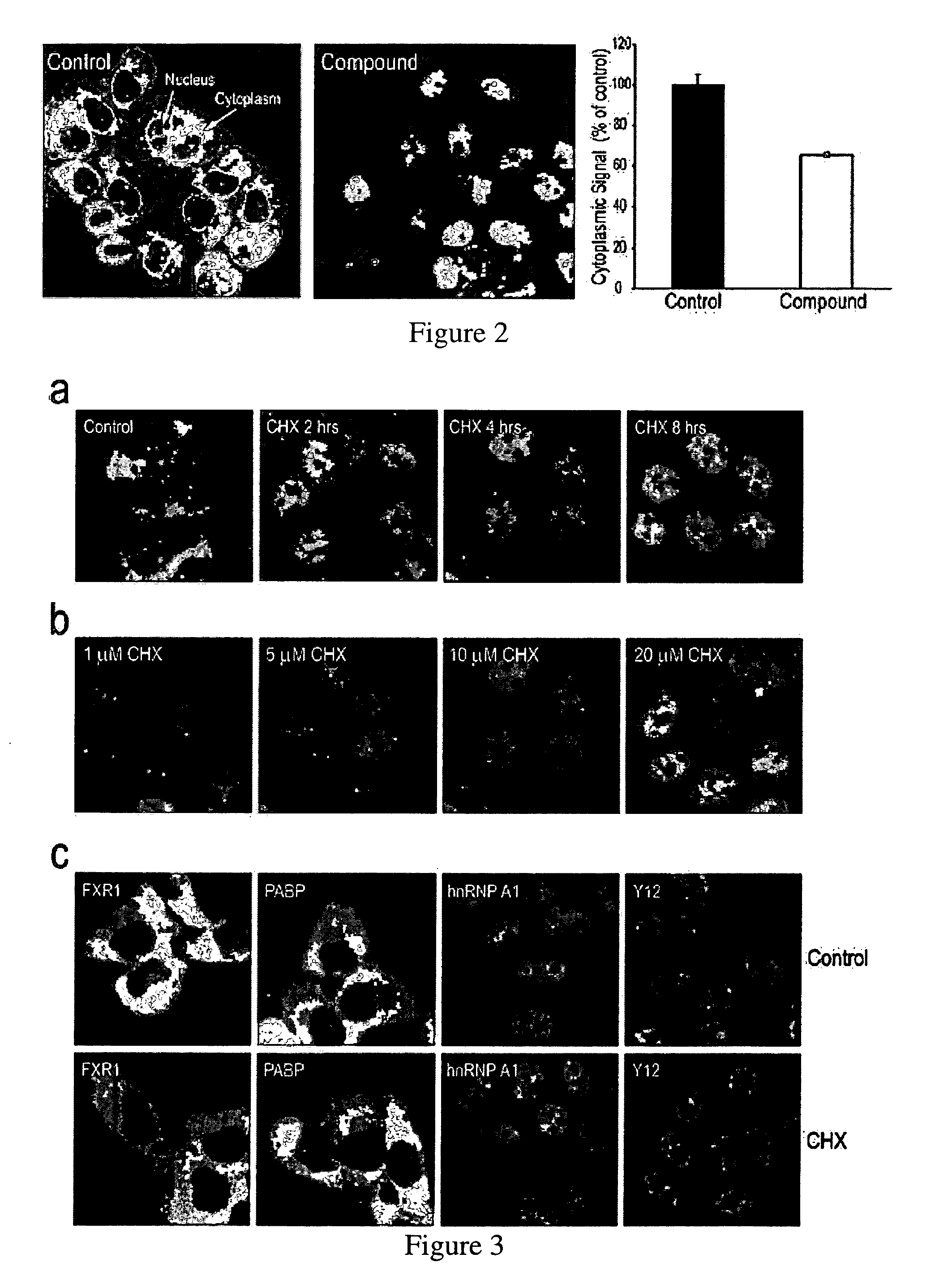
Figure 2 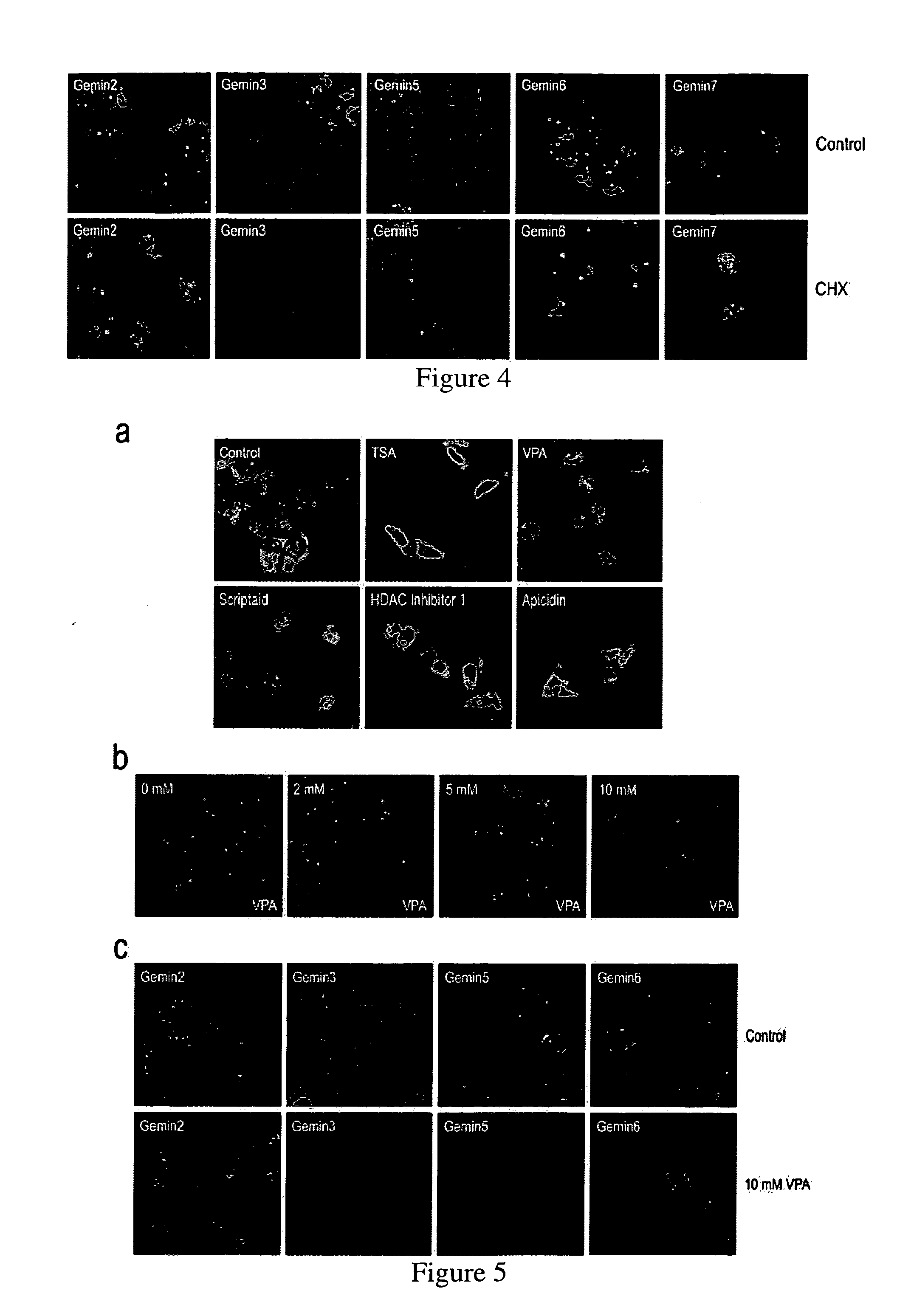
Figure 3
Abstract
Description
FIELD OF INVENTION
[0001] This invention provides: methods, kits, and systems for testing protein synthesis inhibitors.BACKGROUND OF THE INVENTION
[0002] Translation is the RNA directed synthesis of polypeptides. This process requires all three classes of RNA. Although the chemistry of peptide bond formation is relatively simple, the processes leading to the ability to form a peptide bond are exceedingly complex. The template for correct addition of individual amino acids is the mRNA, yet both tRNAs and rRNAs are involved in the process. The tRNAs carry activated amino acids into the ribosome which is composed of rRNA and ribosomal proteins. The ribosome is associated with the mRNA ensuring correct access of activated tRNAs and containing the necessary enzymatic activities to catalyze peptide bond formation.
[0003] Regulation of this information pathway can be achieved at a number of levels, including the modulation of translation factor levels or activity, ribosome biogenesis, and small ...
Examples
example 1
SMN Complex Composition
[0152]In order to determine whether Gems are affected by changes in the composition of the complex, the amount of several Gemins was reduced, one at a time, by RNA interference. The morphology of Gems was monitored by immunofluorescence microscopy using the anti-SMN monoclonal antibody 2B1.
[0153]Reduction of Gemin3 or Gemin4 caused large changes in the size, number and SMN signal intensity of Gems (FIG. 1a). The effect of the treatments was quantified with software analysis of the collected images from a large number of cells. The results indicated a two-fold increase in both signal intensity as well as gem number in each case (FIG. 1b). Thus, changes in the SMN complex are reflected by changes in the morphology of Gems.
example 2
[0154]Based on the results obtained in Example 1, a library of ˜5,000 biologically active small molecules was screened on HeLa cells in 384-well plates. Each well was incubated with a different compound (at 10 μM) and the cells were processed for indirect immunofluorescence. Digital images were collected from six fields in each well and analyzed using imaging algorithms for multiple parameters including relative nuclear and cytoplasmic intensities, and number, size and signal intensity of Gems. DAPI-stained images were collected in a separate channel to define the boundary of each nucleus. Images of individual fields in wells were flagged by the software as showing a significant change in SMN sub-cellular localization. These individual wells were examined directly, and active compounds were re-tested for verification (FIG. 2).
example 3
Redistribution of SMN from the Cytoplasm to the Nucleus
[0155]The previous Examples screened compounds that caused changes in Gems. However, it was noticed that several compounds produced a massive redistribution of SMN from the cytoplasm to the nucleus within a relatively short time (2-6 hours) of treatment (according to the materials and methods of example 2). The most striking accumulation of SMN in the nucleus was observed after treatment with the commonly used protein synthesis inhibitor, cycloheximide (CHX). The effect of CHX was evident after only 2 hours of treatment and there was little SMN detected in the cytoplasm after 8 or more hours of treatment. CHX effect on redistribution of SMN from the cytoplasm to the nucleus was concentration-dependent exhibiting a clear effect even at low micromolar concentrations (0.1-40 μM) (FIG. 3a, b). The nuclear accumulation of SMN did not require complete inhibition of protein synthesis and was proportional to the extent of inhibition of ...