

Partition method of three dimensional medical images based on ray casting volume rendering algorithm
A medical image and volume rendering technology, applied in the field of physical subdivision of three-dimensional medical images, can solve the problems that two-dimensional cannot meet medical applications, and achieve the effects of accurate medical analysis and judgment, and fast image segmentation speed.
Inactive Publication Date: 2009-11-11
UNIV OF ELECTRONICS SCI & TECH OF CHINA
View PDF0 Cites 18 Cited by
- Summary
- Abstract
- Description
- Claims
- Application Information
AI Technical Summary
Problems solved by technology
[0005] However, due to the differences in the imaging principles of medical images and the characteristics of the tissues themselves, and the formation of images is subject to such factors as noise, field offset effects, local body effects and tissue motion, between tissues, between tissues and organs, between organs and organs Due to the influence of space and so on, two-dimensional region segmentation can no longer meet the current medical applications.
Method used
View moreImage
Smart ImageViewing Examples

Examples
Embodiment Construction
[0029] Firstly, simulate on Matlab to obtain the required simulation results, then use C++ language to write the code for reconstructing the 3D model and the code for physical division, and then input the model as source data into the model simplification program in the VC++ interface for processing .
Login to View More PUM
 Abstract
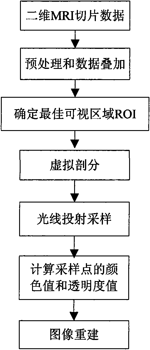
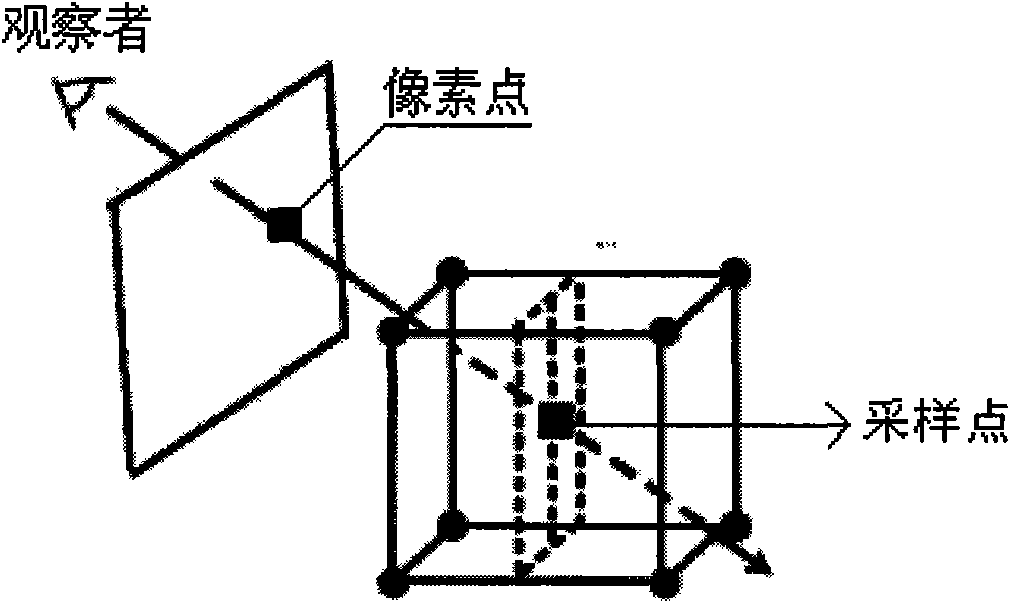
A partition method of three dimensional medical images based on ray casting volume rendering algorithm belongs to the technical field of image processing and relates to the physical partition method of three dimensional medical images. The method includes the following steps of: first converting a two dimensional MRI slice image sequence into three dimensional cuboid data Volume1, revolving the three dimensional cuboid data Volume1 to obtain an optimal visible area ROI; then adopting a regular geometry Volume2 with space size less than that of Volume1 to conduct virtual partition on the Volume1 and to obtain a space geometry Volume3 after the Volume1 is partitioned from the Volume2; subsequently conducting ray casting sampling on the space geometry Volume3; and finally conducting image reconstruction on the space geometry Volume3. Different from a common partition method which first partitions the three dimensional medical images and then reconstructs images, the partition method conducts the partition and three dimensional reconstruction simultaneously, thus increasing arithmetic speed greatly, having high real-time and being capable of observing the required regions at real time through methods of rotation, light illumination and the like so as to find out the association among tissues and facilitate accurate medical analysis and judgment.
Description
technical field [0001] The invention belongs to the technical field of image processing, and relates to a physical subdivision method of three-dimensional medical images. Background technique [0002] In order to accurately distinguish normal tissue structures and abnormal lesions in medical images, medical images need to be segmented. The traditional two-dimensional image segmentation methods mainly include: [0003] (1) Edge-based segmentation methods: usually use different properties between regions (such as gray discontinuity in the region) to divide the boundaries between regions, such methods include parallel differential operator methods (such as Roberts, Sobel, Laplacian, Marr and other operators), serial boundary search methods, methods based on surface fitting, etc.; [0004] (2) Region-based segmentation methods: Usually, the homogeneity within the same region is used to identify different regions in an image, including threshold methods, region growing and spli...
Claims
Login to View More Application Information
Patent Timeline
 Patent Type & AuthorityApplications(China)
IPC IPC(8): G06T7/00G06T15/00
Inventor解梅甄政
OwnerUNIV OF ELECTRONICS SCI & TECH OF CHINA