Sine distortion image correction method used for confocal endoscope

An image correction and endoscopy technology, applied in the field of medical devices, can solve problems such as difficulty in providing subcellular resolution, achieve good application prospects and value, shorten the research and development cycle, and reduce the effect of research and development investment.

Inactive Publication Date: 2018-06-15
EZHOU INST OF IND TECH HUAZHONG UNIV OF SCI & TECH +1
View PDF1 Cites 6 Cited by
  • Summary
  • Abstract
  • Description
  • Claims
  • Application Information

AI Technical Summary

Problems solved by technology

[0005] The invention provides a sinusoidal distortion image correction method for confocal endoscopes, which is used to solve the technical problem that

Method used


Image

  • Sine distortion image correction method used for confocal endoscope
  • Sine distortion image correction method used for confocal endoscope
  • Sine distortion image correction method used for confocal endoscope

Examples


Embodiment 1

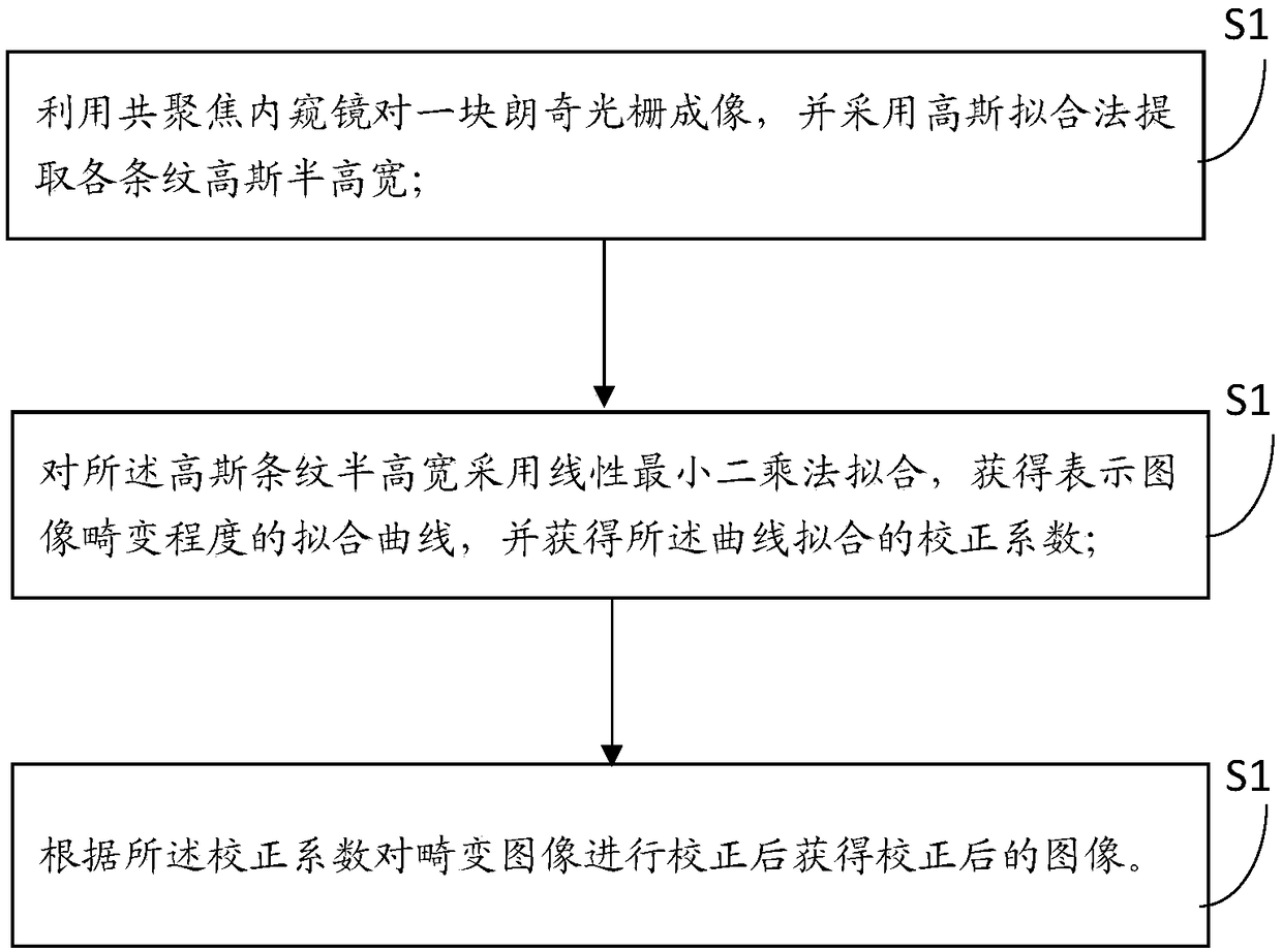
[0029] figure 1 It is a schematic flowchart of a sinusoidal distortion image correction method for a confocal endoscope in an embodiment of the present invention. Such as figure 1 As shown, the method includes:

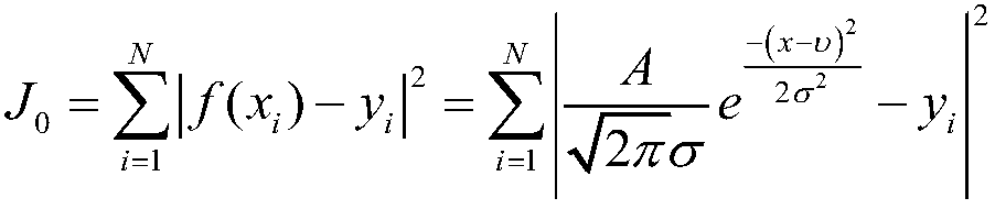
[0030] Step 1: Use a confocal endoscope to image a Ronchi grating, and use the Gaussian fitting method to extract the Gaussian FWHM of each fringe;

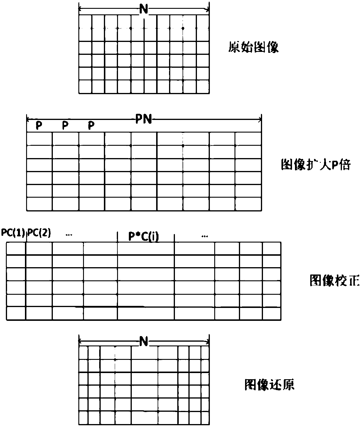
[0031] Further, step 1 also includes: using a confocal endoscope to obtain a grating image after imaging a Ronchi grating; after performing peak detection on the gray value of the first row of the grating image, obtaining the gray value on the first row of the image Points with extremely large grayscale values ​​and points with extremely small grayscale values; Obtain the pixels between two adjacent points with extremely large grayscale values ​​to form a raster stripe; Obtain the points between adjacent two points with extremely small grayscale values The pixels in between form a grating stripe; obtain the pixels in ...


PUM


Abstract

The invention discloses a sine distortion image correction method used for a confocal endoscope, and relates to the technical field of medical instruments. The method comprises the steps that step 1:imaging of a ronchi grating is performed by using the confocal endoscope, and the Gaussian half-height width of each stripe is extracted by using a Gaussian fitting method; step 2: fitting of the Gaussian stripe half-height width is performed by using a linear least square method so as to acquire a fitting curve representing the degree of image distortion and acquire the correction coefficient ofcurve fitting; and step 3: the distortion image is corrected according to the correction coefficient and then the corrected image is acquired. The technical effects that the structural information ofsubcellular level resolution can be provided in an in-vivo way in real time, the method can be used for small animal experiments to shorten the drug research and development period and reduce researchand development input and thus the method has great application prospect and value can be achieved.

Description

technical field [0001] The invention relates to the technical field of medical devices, in particular to a sinusoidal distortion image correction method for a confocal endoscope. Background technique [0002] With the continuous improvement of medical diagnostic methods, the demand for real-time imaging in patients is also increasing. Currently, commonly used equipment such as ultrasound and nuclear magnetic resonance can image human tissues and organs. [0003] However, in the process of realizing the technical solution of the present invention, the inventor of the present application found that the above-mentioned prior art has at least the following technical problems: [0004] The resolutions of commonly used devices in the prior art are all above tens of microns, and it is difficult to provide resolution at the subcellular level. Contents of the invention [0005] The invention provides a sinusoidal distortion image correction method for confocal endoscopes, which is...

Claims


Application Information

Patent Timeline
IPC IPC(8): G06T5/00
CPCG06T5/006
Inventor 刘谦郭琛琛
Owner EZHOU INST OF IND TECH HUAZHONG UNIV OF SCI & TECH
Why Eureka
Try Eureka