Brain tumor image segmentation algorithm based on improved full-convolution neural network

A convolutional neural network and image segmentation technology, applied in the field of medical imaging, can solve the problem of low segmentation accuracy of brain tumor images and achieve strong stability

Inactive Publication Date: 2018-11-27
TIANJIN UNIV
View PDF7 Cites 23 Cited by
  • Summary
  • Abstract
  • Description
  • Claims
  • Application Information

AI Technical Summary

Problems solved by technology

[0007] Aiming at the problem of low accuracy of brain tumor image segmentation by existing machine learning and CNN algorithms, the present invention provides an MR brain tumor image segmentation method based on an improved fully convolutional neural network and a conditional random field. The neural network algorithm FCNN-4s obtains the rough segmentation result, and then uses the statistical learning probability CRF algorithm to correct the brain tumor boundary in the rough segmentation result

Method used


Image

  • Brain tumor image segmentation algorithm based on improved full-convolution neural network
  • Brain tumor image segmentation algorithm based on improved full-convolution neural network
  • Brain tumor image segmentation algorithm based on improved full-convolution neural network

Examples


Embodiment Construction

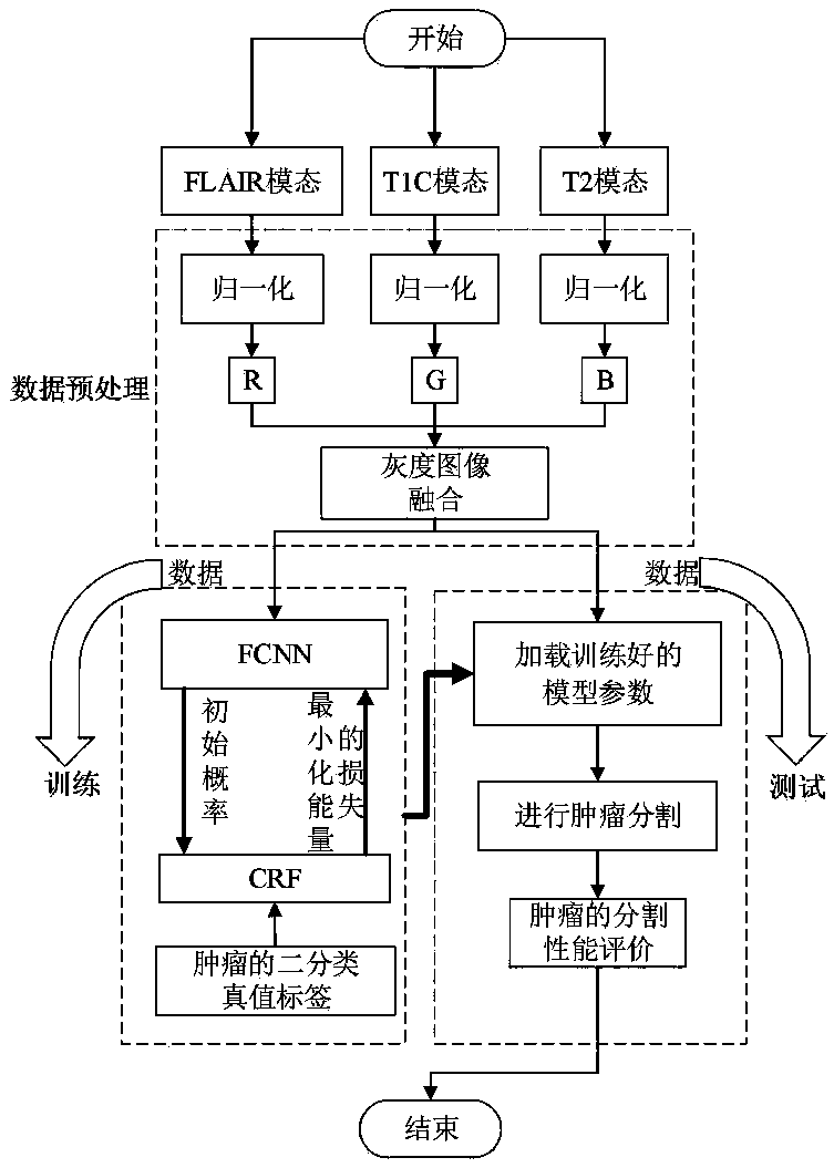
[0031] The brain tumor image segmentation algorithm process of the improved fully convolutional neural network of the present invention is as follows: figure 1shown. Firstly, grayscale image fusion is performed on the normalized multimodal MR brain tumor image data; then part of the fused data is used as a training set, and part of the data is used as a test set, and the training set is used to train the fusion algorithm of FCNN and CRF; finally, in the test The test of the model and the index evaluation of the segmentation results are carried out on the set.

[0032] 1) Image preprocessing

[0033] Each pixel of the original MR brain tumor image is 16bit, but in the digital image processing technology, the 8bit image is processed, so the present invention first normalizes the grayscale of the MR image, and the grayscale of each pixel is Degree values ​​are uniformly compressed to a range between 0-255. Considering that the features extracted by the neural network only thro...


PUM


Abstract

The invention relates to a MR brain tumor image segmentation method based on an improved full-convolution neural network algorithm. The method comprises the following steps: step 1, image preprocessing; step 2, improved FCNN coarse segmentation algorithm, including based on a FCNN, adding a batch regularization layer after each convolution layer to speed up training of the network and improve precision of a model, and performing features fusion on pooled brain tumors features for three times, to obtain more refined brain tumors features, and establishing an improved FCNN, called a FCNN-4s network; step 3, FCNN-4s and CRF resegmentation fusion algorithm, including initializing an energy function in a CRF model according to a FCNN-4s coarse segmentation result in the second step, to obtain aprobability value of pixel original home label, and then calculating the CRF model by the following steps, continuously iterating and correcting two kinds of probability maps of FCNN-4s prediction, to obtain a CRF resegmentation fusion result.

Description

technical field [0001] The invention is an important field in the field of medical imaging, and combines medical images with deep learning algorithms to complete accurate segmentation of brain tumor nuclear magnetic resonance images. Background technique [0002] Malignant brain tumors are one of the most feared types of cancer in the world, often leaving patients with cognitive decline and poor quality of life. The most common brain tumors in adults are primary central nervous system lymphoma and glioma, among which glioma accounts for more than 80% of malignant tumors, so glioma is the key object of tumor segmentation. However, since gliomas can appear anywhere in the brain with variable sizes and irregular shapes, their segmentation is still a challenging task. Therefore, how to segment brain tumors efficiently and fully automatically using modern information technology has become an important issue. research direction. Magnetic Resonance Imaging (MRI) technology is non...

Claims


Application Information

Patent Timeline
Patent Type & Authority Applications(China)
IPC IPC(8): G06K9/34G06K9/62G06N3/04
CPCG06V10/267G06N3/045G06F18/2193G06F18/253
Inventor 邢波涛李锵关欣
Owner TIANJIN UNIV
Why Eureka
Try Eureka