Rapid imaging method, device and equipment for nuclear magnetic resonance image

A technology of nuclear magnetic resonance images and imaging methods, applied in the field of image processing, can solve the problems of long MR image time and low MR image prediction accuracy, and achieve the effects of reducing training time, accelerating convergence speed, and improving initialization speed.

A technology of nuclear magnetic resonance images and imaging methods, applied in the field of image processing, can solve the problems of long MR image time and low MR image prediction accuracy, and achieve the effects of reducing training time, accelerating convergence speed, and improving initialization speed.

CN109801259AInactive Publication Date: 2019-05-24SHENZHEN INST OF ADVANCED TECH CHINESE ACAD OF SCI

Image

  • Rapid imaging method, device and equipment for nuclear magnetic resonance image
  • Rapid imaging method, device and equipment for nuclear magnetic resonance image
  • Rapid imaging method, device and equipment for nuclear magnetic resonance image

Examples


Embodiment Construction

[0045] In the following description, specific details such as specific system structures and technologies are presented for the purpose of illustration rather than limitation, so as to thoroughly understand the embodiments of the present application. It will be apparent, however, to one skilled in the art that the present application may be practiced in other embodiments without these specific details. In other instances, detailed descriptions of well-known systems, devices, circuits, and methods are omitted so as not to obscure the description of the present application with unnecessary detail.

[0046] In order to illustrate the technical solutions described in this application, specific examples are used below to illustrate.

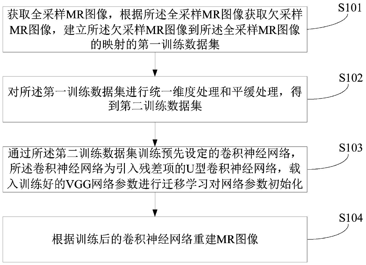
[0047] figure 1 It is a schematic diagram of the implementation flow of a fast imaging method for nuclear magnetic resonance images provided in the embodiment of the present application, and is described in detail as follows:

[0048] In step S101, a ...


PUM


Abstract

A rapid imaging method of the nuclear magnetic resonance image comprises the steps of obtaining a full-sampling MR image, obtaining an under-sampling MR image according to the full-sampling MR image,and establishing a first training data set of mapping from the under-sampling MR image to the full-sampling MR image; Performing unified dimension processing and smooth processing on the first training data set to obtain a second training data set; A preset convolutional neural network is trained through the second training data set, the convolutional neural network is a U-shaped convolutional neural network with introduced residual terms, and trained VGG network parameters are loaded for transfer learning to initialize the network parameters; And reconstructing an MR image according to the trained convolutional neural network. The accuracy of network classification can be effectively improved, the initialization speed of network parameters is reduced, and the training efficiency of the network is improved.

Description

technical field [0001] The present application belongs to the field of image processing, and in particular relates to a fast imaging method, device and equipment for nuclear magnetic resonance images. Background technique [0002] Magnetic resonance imaging (MRI) has been applied to the imaging diagnosis of various systems of the whole body. It can display fat, whole body organs, muscles, fast-flowing blood, bones and air, etc., and can also clearly display the internal structure of organs. For cardiovascular diseases, it can not only observe the anatomical changes of chambers, great blood vessels and valves, but also can be used for ventricle analysis for qualitative and semi-quantitative diagnosis. It can make multiple slices with high spatial resolution to display the heart and lesions The overall picture and its relationship with the surrounding structures are superior to other X-ray imaging, two-dimensional ultrasound, nuclide and CT examinations. Doctors can very well...

Claims


Application Information

Patent Timeline
24 May 2019
Publication
CN109801259A
IPC
G06T7/00
Inventors
杨洁; 李文