Histopathology image classification method based on color deconvolution and self-attention model

A technology of attention model and classification method, applied in the field of tissue and cell image classification combining color deconvolution and self-attention model, can solve the problems of low efficiency, low accuracy, cumbersome steps, etc., and achieve the effect of good classification effect

Pending Publication Date: 2022-02-18
FUJIAN NORMAL UNIV
View PDF0 Cites 2 Cited by
  • Summary
  • Abstract
  • Description
  • Claims
  • Application Information

AI Technical Summary

Problems solved by technology

[0004] In the past, the methods of manually extracting image features or using machine learning were often used for the classification of histopathological images. However, these methods have the characteristics of cumbersome steps, low efficiency, and low accuracy, and are difficult to use in the actual tissue cell image classification process.

Method used


Image

  • Histopathology image classification method based on color deconvolution and self-attention model
  • Histopathology image classification method based on color deconvolution and self-attention model
  • Histopathology image classification method based on color deconvolution and self-attention model

Examples


Embodiment Construction

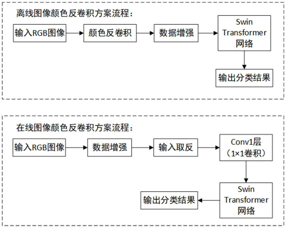
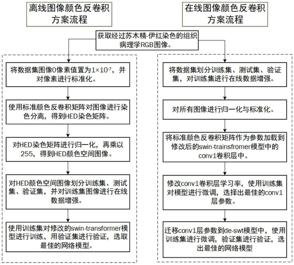
[0066] see Figure 1-Figure 2 As shown in one of the embodiments, the embodiment includes two implementation methods of offline image color deconvolution or online image color deconvolution.

[0067] Offline image color deconvolution implementation method:

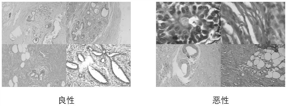
[0068] see image 3 As shown, in S100, obtain a histopathological image data set stained with hematoxylin-eosin. In this embodiment, the BreakHis breast cancer histopathological image data set is selected. The data set consists of 7909 images collected from 82 patients. Among them, there were 24 benign patients, from which 2480 benign images were collected; 58 malignant patients, from which 5429 malignant images were collected, benign image and malignant image samples. There are four magnifications in the dataset, 40x, 100x, 200x, and 400x. The image size is 700×460, and it is scaled to a 224×224 size image, and the pixels of each image form a matrix H with a size of 224×224×3 t , where 3 corresponds to the R, G, and B...


PUM


Abstract

The invention relates to a histopathology image classification method based on color deconvolution and a self-attention model, and provides an offline image color deconvolution scheme and an online image color deconvolution scheme, and the offline image color deconvolution scheme comprises the following steps: firstly, carrying out deconvolution on an RGB image to obtain an HED color space image; inputting the HED image into a self-attention model to obtain a classification result of the image; according to the online color deconvolution scheme, deconvolution operation is added into a self-attention model in a convolutional layer form, the model is trained and proper deconvolution parameters are searched, then the deconvolution parameters are migrated, and the self-attention model added with the deconvolution operation is utilized again to classify images; by adopting the technical scheme, the histopathology image deconvolution and the self-attention model are effectively combined, and the accuracy of histopathology image classification is improved.

Description

technical field [0001] The invention belongs to the field of medical tissue cell image processing, in particular to a tissue cell image classification method combining color deconvolution and a self-attention model. Background technique [0002] According to data released by the International Agency for Research on Cancer of the World Health Organization, in 2020, there will be about 19.3 million new cancer cases and about 10 million deaths worldwide. Among them, the number of new breast cancer cases reached 2.26 million, accounting for 11.7% of the global new cancer cases, surpassing the 2.2 million new cases of lung cancer, becoming the world's largest cancer. Examination of breast tissue sections by pathologists remains the gold standard for clinical diagnosis during breast cancer diagnosis. With the development of digital pathology, the number of digital tissue cell images has exploded. With the improvement of people's health awareness and the gradual popularization of ...

Claims


Application Information

Patent Timeline
Patent Type & Authority Applications(China)
IPC IPC(8): G06T7/00G06T7/90G06K9/62G06V10/764G06V10/774G06N3/04G06N3/08
CPCG06T7/0012G06T7/90G06N3/04G06N3/084G06T2207/10056G06T2207/20081G06T2207/20084G06T2207/30096G06F18/24G06F18/214
Inventor 何柱林铭炜钟美荟姚志强
Owner FUJIAN NORMAL UNIV
Why Eureka
Try Eureka