Three dimensionally orientated cardiac PCT-CT and MR1 image imersing diagnosis and apparatus

A PET-CT and stereotaxic technology, applied in the field of medical imaging examination, can solve the impossible and inability to achieve precise positioning scanning and other problems, and achieve the effect of high application value

Inactive Publication Date: 2005-07-27
GENERAL HOSPITAL OF TIANJIN MEDICAL UNIV
View PDF0 Cites 5 Cited by
  • Summary
  • Abstract
  • Description
  • Claims
  • Application Information

AI Technical Summary

Problems solved by technology

At present, since PET-CT and MRI detection are carried out separately, it is impossible to accurately locate the scan, so as to obtain the detection results at the same l...

Method used


Image

  • Three dimensionally orientated cardiac PCT-CT and MR1 image imersing diagnosis and apparatus

Examples


Embodiment Construction

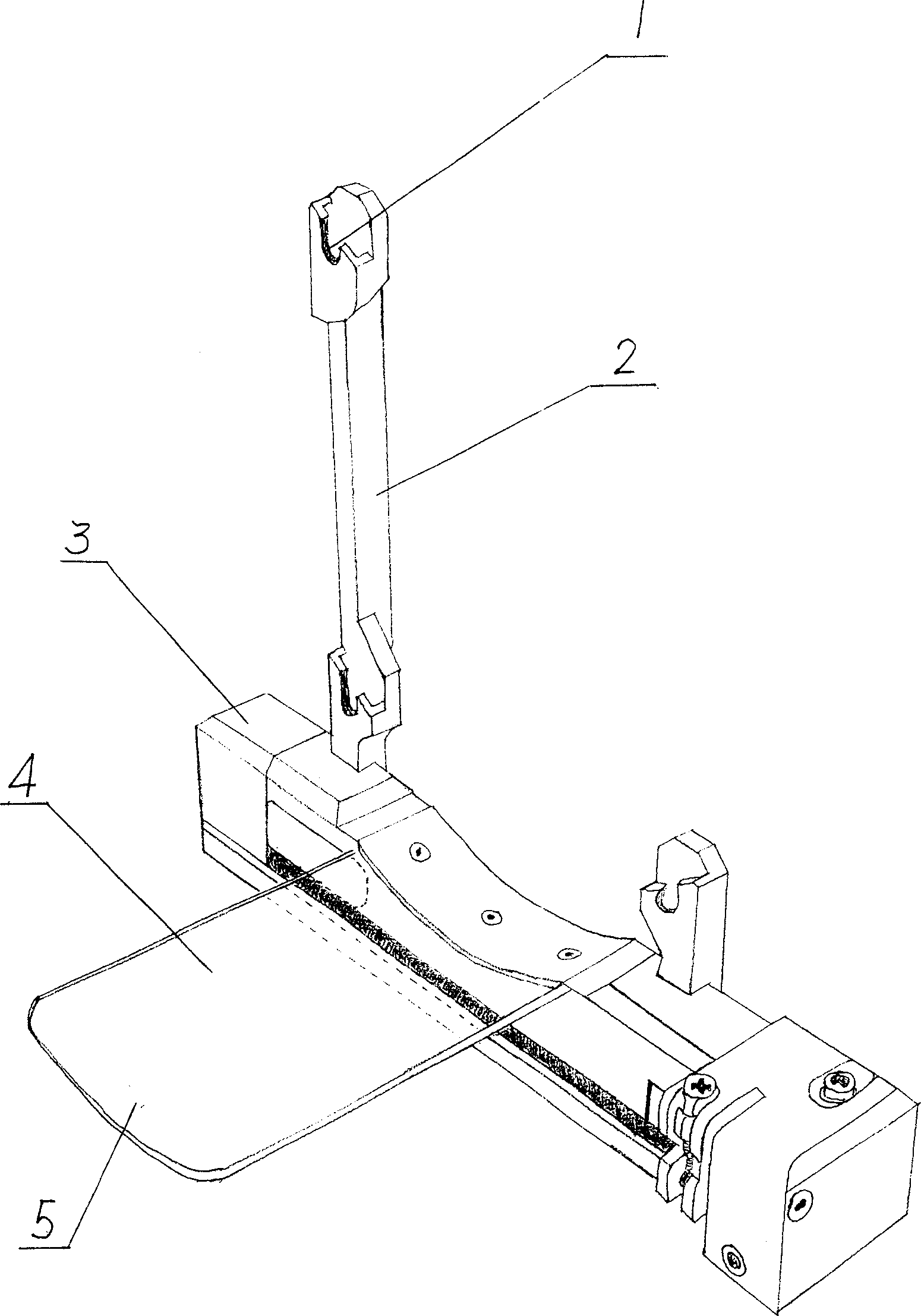
[0026] The embodiment of the present invention is based on using the Leksell stereotaxic system produced by Sweden's Elekta Company to fix a stereotaxic frame on the patient's head and insert a positioning plate, and then perform PET-CT and MRI, or MRI and PET-CT scanning. Then input the MRI scan image information and PET-CT scan image information into the computer, integrate and reconstruct the PET-CT scan image information on the MRI scan image at the same level through Photoshop computer software, and then use different colors to mark each Abnormal areas of the image were identified.

[0027] During the MRI scan, the patient lies supine on the magnetic resonance MRI examination table, the Leksell stereotaxic frame is fixed by the connector on the MRI examination table, and then the MRI axial scan is performed along the baseline of the stereotaxic frame. Choose different sequences or enhanced scans according to the purpose of detection or treatment, which can be plain scans ...


PUM


Abstract

A method and device for stereoscopically and directionally examining the PET-CT and MRI images of skull at same time by combining them together is disclosed. The same layer of skull for PET-CT and MRI images can be precisely repeated and combined for processing, so directly and really showing the lesion in skull.

Description

【Technical field】 [0001] The invention relates to medical imaging examination, in particular to a stereotactic head PET-CT and MRI image fusion examination method and device. 【Background technique】 [0002] With the development of modern science and technology, advanced computerized tomography (Computed Tomography, CT) came out in the 1970s, using a method completely different from ordinary X-ray photography, which can display tomography of various tissues and organs in the body. The development of neuroimaging ushered in a new era. The emergence and application of Magnetic Resonance Image (MRI) in the 1980s was even more icing on the cake in the field of imaging. It has the advantages of hydrogen ion imaging, multi-sequence, three-dimensional, high resolution of tissue structure, and non-radiation (such as 1.5T Twin Speed ​​infinity with Excite I superconducting magnetic resonance imaging system produced by American GE company). They can perform special depiction on vascu...

Claims


Application Information

Patent Timeline
IPC IPC(8): A61B5/055A61B6/03
Inventor 杨卫东孙健高硕曾增蔡丽刘国栋李富华
Owner GENERAL HOSPITAL OF TIANJIN MEDICAL UNIV
Why Eureka
Try Eureka