Visualizing ablation cannula

a cannula and visualization technology, applied in the field of medical devices and procedures, can solve the problems of lbp being the most expensive of all medical diagnoses, pain originating in the facet joints, and a large portion of the procedure is quite uncomfortable, so as to reduce the origin of lbp, eliminate or minimize, and precise tailor the effect of laser treatmen

Active Publication Date: 2005-08-18
ARDENT MEDICAL CORP
View PDF13 Cites 17 Cited by
  • Summary
  • Abstract
  • Description
  • Claims
  • Application Information

AI Technical Summary

Benefits of technology

[0011] The cannula of the needle set can be used for placement on a tissue site in a subject's anatomy. The cannula can be hollow and can define a longitudinal channel or lumen. The distal tip of the cannula can include a tissue-gripping surface to prevent slippage of the cannula, once positioned at the target site. The needle set can also include a removable trocar for occluding the lumen of the cannula during placement of the cannula at a tissue site within the subject's anatomy. In one aspect of the present invention, the trocar can be a tubular shaft having a pointed distal tip.
[0013] The invention further encompasses a method of using the visualizing tissue ablation needle set in a surgical procedure for endoscopic laser ablation of tissues. In an exemplary use of this procedure, a surgeon may achieve denervation of the pain-causing nerve fibers of the facet joints, thereby reducing LBP originating from the facet joints. The method allows for visualization of the facet joint tissue prior to and during application of a laser beam used for ablation of the nerves innervating the joint.
[0014] The advantages of incorporating an endoscopic tool into a tissue ablation needle set are several. After initial placement of the cannula with radiographic guidance, and following insertion of the visualizing ablation probe into the cannula, anatomical structures at the tip of the probe can be visualized on the screen of the endoscope. Accordingly, the accuracy of the initial needle placement with respect to the target tissue can be assessed directly, allowing for corrections of the needle position. This advantage eliminates or minimizes the need for repeated insertions of the probe near the target site. Similarly, during the ablation procedure performed with the laser tool, the clinician is able to directly visualize the extent of the laser lesioning during the application of the laser treatment, allowing for precise tailoring of the laser treatment according to the response of the particular subject's tissues.
[0015] A further advantage of a needle set having a cannula with a tissue-gripping surface is that the position of the cannula can be anchored on solid anatomical structures such as bone and fibrous tissue forming joint capsules. By gripping onto the bone or fibrous tissue, the tissue-gripping surface can prevent slipping of the cannula tip away from the intended site of treatment. This feature, particularly in conjunction with the ability to view the site of the target tissue by endoscopy, enables the practitioner to stabilize the ablation probe in the precise location of the target, once the target is located.

Problems solved by technology

As the most frequently reported work-related injury, LBP is the most costly of all medical diagnoses when the costs of time lost at work, long-term disability and medical and legal expenses are factored in.
Inflammation, injury, nerve entrapment and degenerative osteoarthritic changes in the joint tissues all can lead to pain originating in the facet joints.
This portion of the procedure is quite uncomfortable and calls for judicious use of sedation and analgesics.
In fact, proper placement of the needle tip in the complicated structure of a subject's spine requires great skill by the treating clinician.
The needles may need to be withdrawn and re-inserted multiple times. Errors in needle placement can result in accidental impalement of structures such as the nerve root in the lower spine, presenting a serious medical risk to the subject.
This wide variability in the procedure in the hands of different practitioners suggests unpredictability inherent in the procedure itself.
The unpredictability may be a reflection of failure of existing methods to enable sufficiently precise localization of the target nerve prior to lesioning, combined with incomplete destruction of the pain-causing nerve fibers by the RF electrode.

Method used


Image

  • Visualizing ablation cannula
  • Visualizing ablation cannula
  • Visualizing ablation cannula

Examples


Embodiment Construction

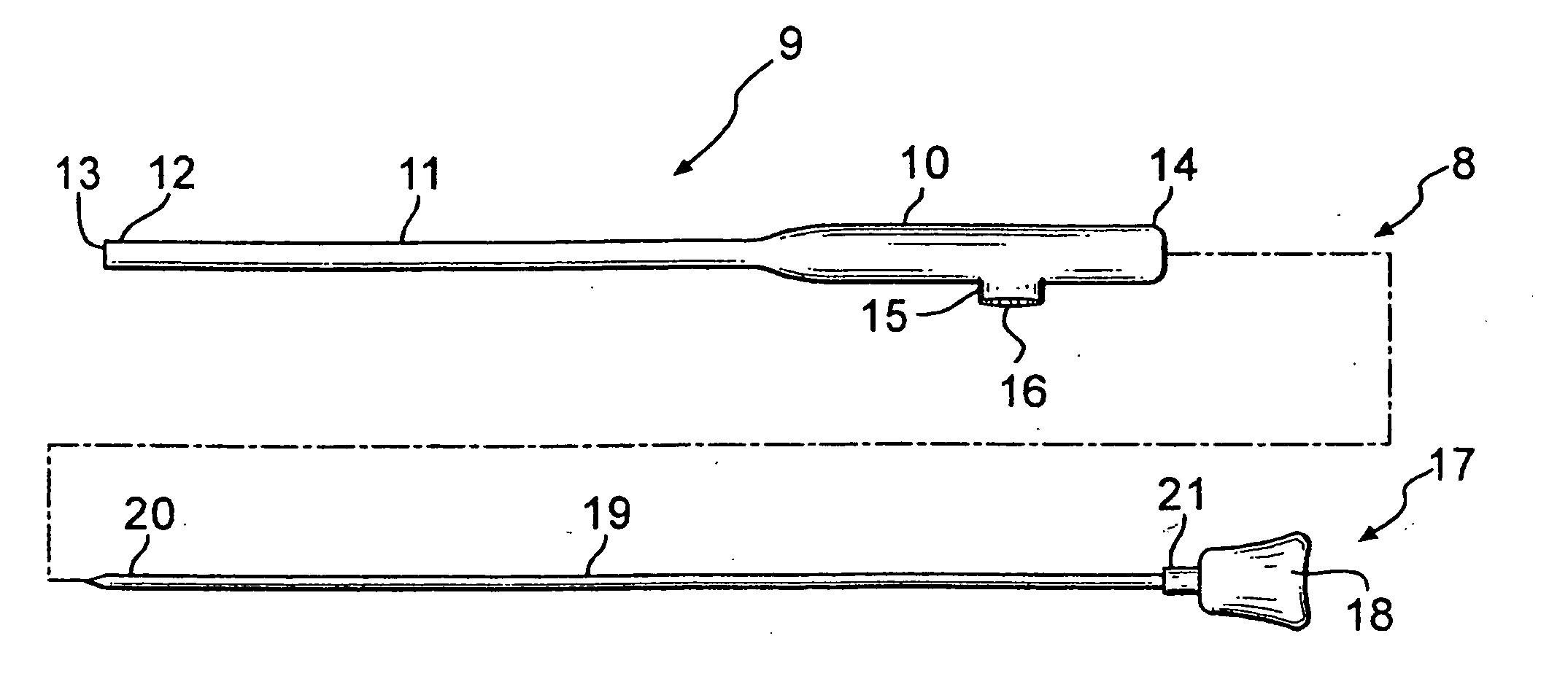
[0022] The present invention is a medical apparatus and surgical procedure for the visualization and ablation of tissue in a subject's body. The apparatus can include a needle set, the needles set including an outer cannula, a trocar and a visualizing ablation probe. Whereas the cannula can be placed at a tissue site requiring treatment, the trocar can be included within the cannula and can be used to occlude the cannula during placement. Once the cannula has been positioned within the subject's body proximate to the target tissue, the visualizing ablation probe can be inserted into the cannula and used to observe and ablate the target tissue during treatment with a medical laser.

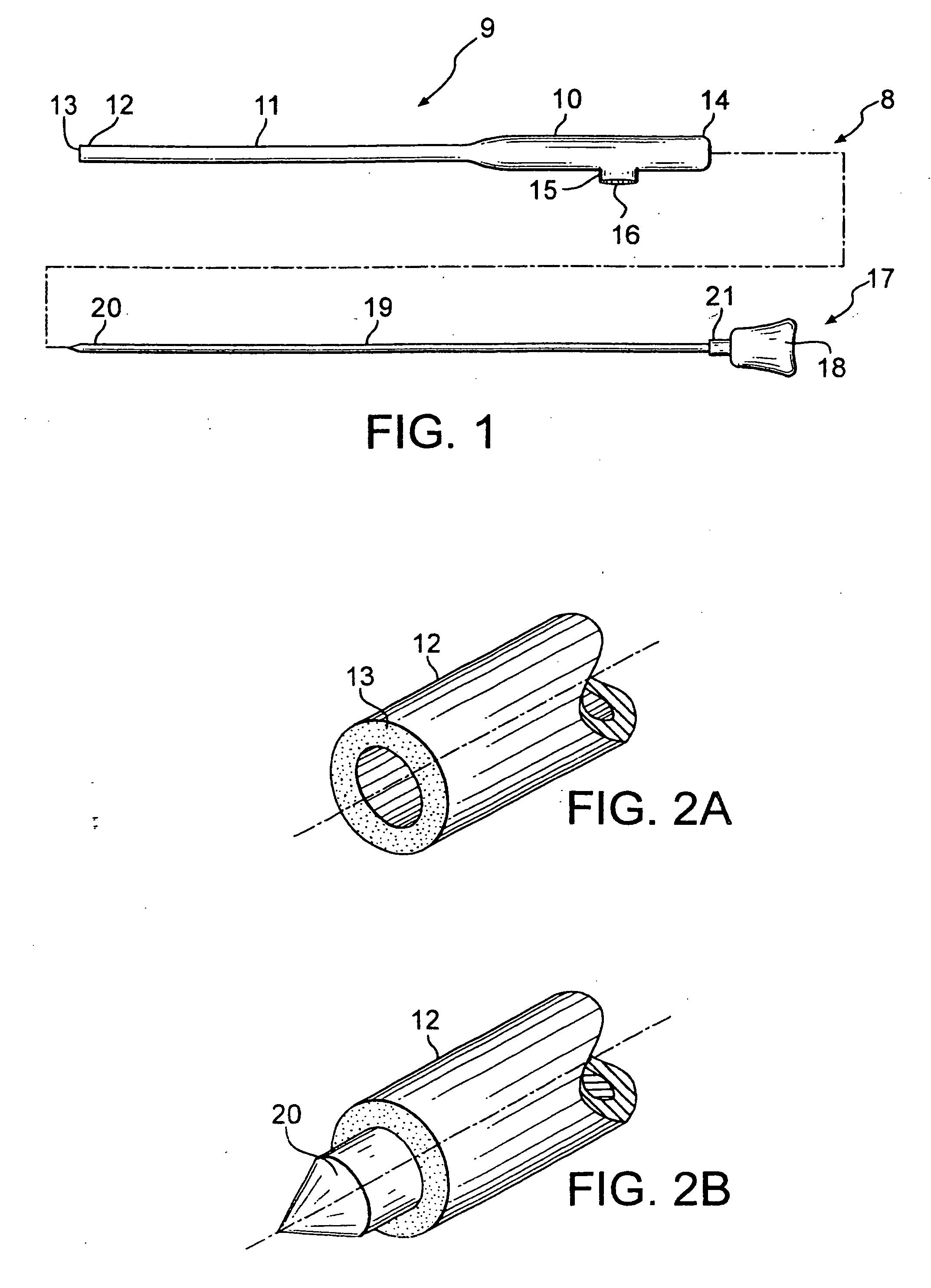
[0023] Referring initially to FIG. 1, several features of the needle set of the invention are shown, including an embodiment of the cannula 8 with a distal tip 12 having a tissue-gripping surface 13. FIG. 1 also depicts an embodiment of the trocar 17 with a pointed tip 20. In the illustrated embodiment of ...


PUM


Abstract

A medical needle set for visualized tissue ablation within a subject's body includes a cannula and components configured for inclusion in the cannula, including a trocar for occlusion of the cannula lumen during needle placement, and a visualizing ablation probe used for simultaneous endoscopic viewing and ablation of tissue sites with a laser beam. The cannula can include a tissue-gripping surface for stabilization of the needle set on the target tissue. A surgical system for tissue ablation includes a visualizing ablation needle set operably connected to an endoscope and a laser. A surgical procedure using this system permits simultaneous visualization and ablation of tissues, including those of the facet joints of the spine.

Description

BACKGROUND OF THE INVENTION [0001] 1. Statement of the Technical Field [0002] The disclosed invention is in the field of medical devices and procedures. In particular, the disclosed invention pertains to a novel surgical device and its use for ablation of tissues associated with solid anatomical surfaces such as joints. [0003] 2. Description of the Related Art [0004] Lower back pain (LBP) is a common musculoskeletal complaint of industrialized society with a reported 60-90% of the population experiencing at least one episode of LPB per lifetime. As such, LBP is a very common cause of disability in persons younger than 45 years, the second leading reason for visits to primary care physicians, and the most frequent cause of visits to orthopedic surgeons and neurosurgeons. As the most frequently reported work-related injury, LBP is the most costly of all medical diagnoses when the costs of time lost at work, long-term disability and medical and legal expenses are factored in. Over the ...

Claims


Application Information

Patent Timeline
Patent Type & Authority Applications(United States)
IPC IPC(8): A61B1/04A61B1/313A61B18/00A61B18/24
CPCA61B1/042A61B2018/00982A61B18/24A61B1/313
Inventor KATZMAN, SCOTT S.
Owner ARDENT MEDICAL CORP
Why Eureka
Try Eureka