Check patentability & draft patents in minutes with Patsnap Eureka AI!

Method for establishing an overlay image to be displayed, display device, computer program, and data medium

a technology of overlay image and display device, applied in image enhancement, instruments, applications, etc., can solve the problems of blurred image information, sharp differences in contrast within the displayed blood vessel system, and further challenges and even restrictions, so as to improve the visibility of individual image information, increase the global contrast of the display, and reduce the effect of restrictions on the ability to make settings

Active Publication Date: 2018-05-03
SIEMENS HEALTHCARE GMBH
View PDF4 Cites 4 Cited by
  • Summary
  • Abstract
  • Description
  • Claims
  • Application Information

AI Technical Summary

Benefits of technology

The patent discusses a method to improve the visibility of medical images in an overlay image. The method involves modifying the image value of one dataset based on the image value of another dataset to enhance the contrast between them. This improves the visibility of the image information and reduces the interaction between the user and the display device. In one embodiment, a linear or non-linear weighting function is applied to the modified dataset, which amplifies the image value in regions of high image value of the base dataset. The method allows the user to set the modification parameters within a range, which further restricts the ability to make settings. Additionally, the method preserves the color coding in the modified image dataset, allowing for the use of color-coded medical image datasets in the overlay image.

Problems solved by technology

The display of a number of items of image information from different image sources in a single overlay image leads to further challenges and even to restrictions.
Individual vessels are not clearly shown here, so this may already result in sharp differences in contrast within the displayed blood vessel system.
If these two medical image datasets are now overlaid, the instrument may be only recognized with great difficulty in very bright vessel regions, which may lead to a misinterpretation of the overlay image.
Similar problems also arise in other application areas, for example, if one of the medical image datasets exploits the available intensity dynamics of the display to the maximum for example and thus the result may display problems at specific contrasts of the other medical image dataset.

Method used


Image

  • Method for establishing an overlay image to be displayed, display device, computer program, and data medium
  • Method for establishing an overlay image to be displayed, display device, computer program, and data medium
  • Method for establishing an overlay image to be displayed, display device, computer program, and data medium

Examples


Embodiment Construction

[0037]The method is to be presented below in an application for image support during a minimally-invasive intervention in a blood vessel system of a patient, in particular, a roadmap procedure. This application is to be understood as an example; the process may also be used above and beyond this in diverse ways, whenever overlay images are to be viewed in a medical context.

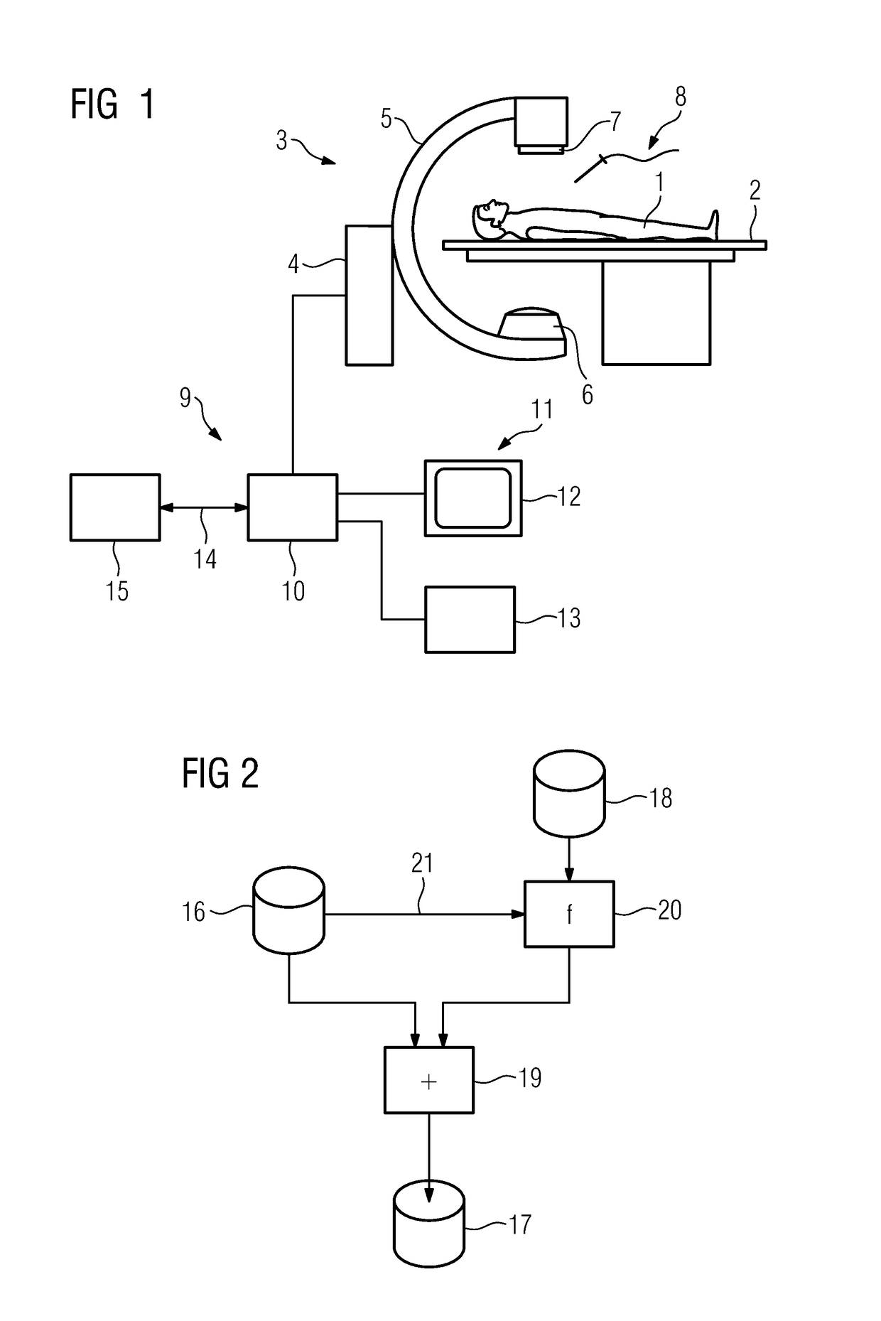
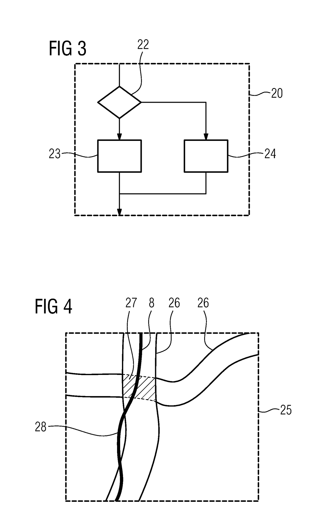
[0038]FIG. 1 depicts a system in which the medical intervention and also the image monitoring may be carried out. The patient 1 to be treated or to be examined is supported in this case on a patient couch 2. Image data of the patient 1 may be recorded with an imaging device 3, here an X-ray device 4 with a C-arm 5, on which an X-ray emitter 6 and an X-ray detector 7 are arranged opposite one another. The minimally-invasive intervention itself is carried out with at least one medical instrument 8, for example, a guide wire and / or a catheter.

[0039]For planning of the minimally-invasive intervention here the X-ray de...


PUM


Abstract

A method is provided for establishing an overlay image to be displayed from medical image datasets of a recording region of a patient registered with one another containing at least two items of different image information. The method includes establishing, for at least a part of the overlay image, an image value of the overlay image at an image position by addition or subtraction of an image value of at least one base image dataset of the medical image datasets at the image position and a modified image value of at least one modification image dataset of the medical image datasets at the image position dependent on the image value of the base image dataset at the image position.

Description

[0001]The application claims the benefit of German Patent Application No. DE 10 2016 221 220.6, filed Oct. 27, 2016, incorporated by reference in its entirety.TECHNICAL FIELD[0002]The disclosure relates to a method for establishing an overlay image to be displayed from medical image datasets of a recording region of a patient registered with one another containing at least two different items of image information. In addition, the disclosure relates to a display device, to a computer program, and to an electronically-readable data medium.BACKGROUND[0003]For a plurality of medical applications, (e.g., in medical diagnostics or in medical therapy), the use of a great diversity of imaging modalities is widely known. In these applications, such diagnostics or such a therapy may be undertaken in many cases using a single medical image dataset, e.g., a single computed tomography (CT) image dataset or a magnetic resonance (MR) image dataset.[0004]Most notably with more complex problems, bu...

Claims


Application Information

Patent Timeline
Patent Type & Authority Applications(United States)
IPC IPC(8): G06T7/33G06T7/00G06T5/00G06T5/20A61B34/20A61B90/00
CPCG06T7/33G06T7/0012G06T5/002G06T5/008G06T5/20A61B34/20A61B90/37G06T2207/10081G06T2207/10104G06T2207/10108G06T2207/10116G06T2207/20182G06T2207/20221G06T2207/30101A61B2090/365A61B2090/374A61B2090/3762A61B2090/378A61B2034/2065G06T5/50G06T2207/10072G06T2207/20012G06T2207/20224A61B2090/364A61B2090/376G06T5/70G06T5/94
Inventor ERTEL, DIRK
Owner SIEMENS HEALTHCARE GMBH
Features
  • R&D
  • Intellectual Property
  • Life Sciences
  • Materials
  • Tech Scout
  • Unparalleled Data Quality
  • Higher Quality Content
  • 60% Fewer Hallucinations
Learn More