

Method for converting tone of chest x-ray image, storage medium, image tone conversion apparatus, server apparatus, and conversion method
a chest x-ray and image technology, applied in the field of processing medical images, can solve the problems of difficult interpretation and unconsidered computer tomography, and achieve the effect of improving the contrast level
- Summary
- Abstract
- Description
- Claims
- Application Information
AI Technical Summary
Benefits of technology
Problems solved by technology
Method used
Image

Examples
embodiments
[0105]Embodiments of the present disclosure will be described hereinafter with reference to the drawings. In the drawings, the same components are given the same reference numerals, and redundant description thereof is omitted as necessary.
first embodiment
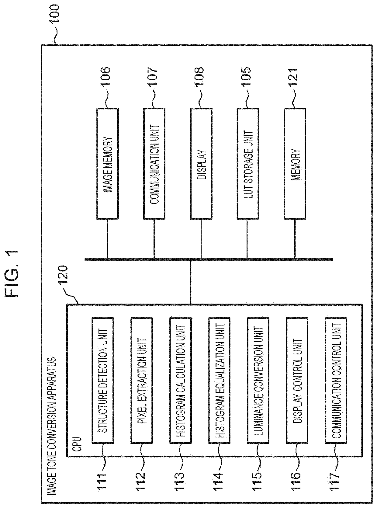
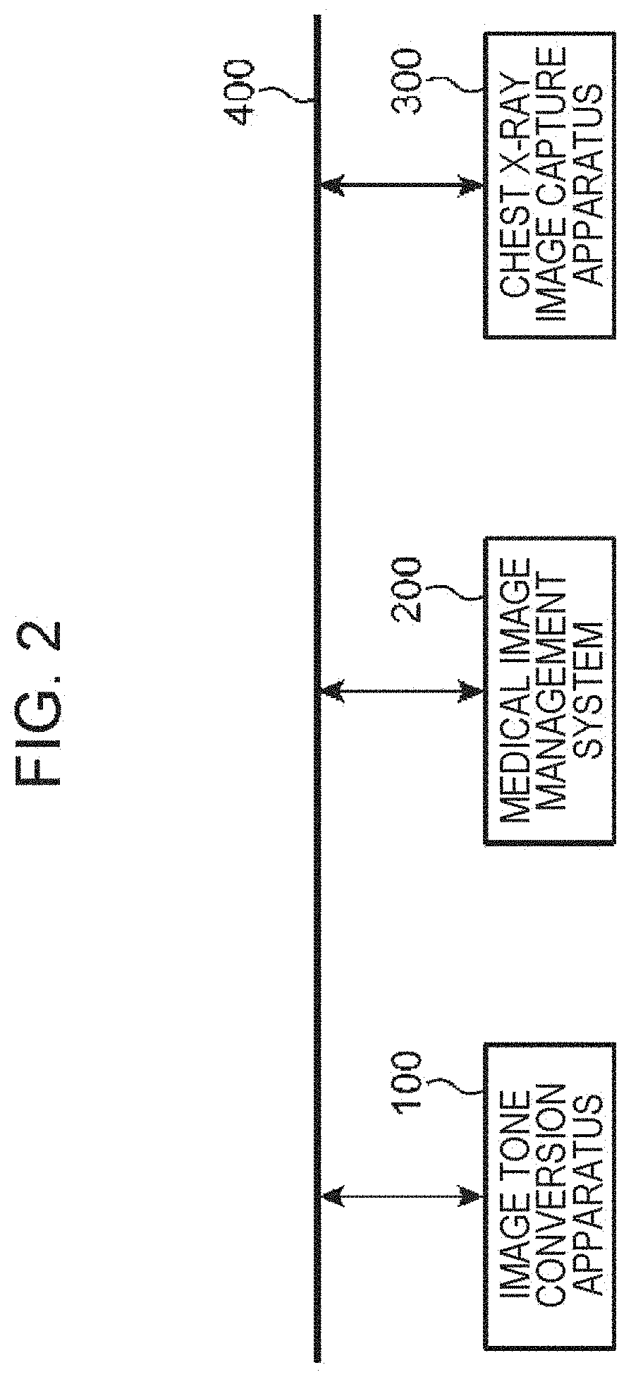
[0106]FIG. 1 is a block diagram schematically illustrating the configuration of an image tone conversion apparatus 100 that performs a method for converting tone of a chest X-ray image according to a first embodiment. FIG. 2 is a block diagram schematically illustrating a network configuration 410 in a medical facility,
[0107]As illustrated in FIG. 2, the network configuration 410 in the medical facility includes an intra network 400. The image tone conversion apparatus 100, a medical image management system 200, and a chest X-ray image capture apparatus 300 are connected to the intra network 400. The medical image management system 200 stores and manages chest X-ray images, computer tomography (CT) images, magnetic resonance imaging (MRI) images, and the like. The chest X-ray image obtaining apparatus 300 captures chest X-ray images of patients and persons who receive a medical examination. Chest X-ray images captured by the chest X-ray image capture apparatus 300 are transmitted an...
second embodiment
[0151]FIG. 12 is a block diagram schematically illustrating the configuration of an image tone conversion apparatus 100A that performs a method for converting tone of a chest X-ray image according to a second embodiment. Unlike the image tone conversion apparatus 100 illustrated in FIG. 1, the image tone conversion apparatus 100A illustrated in FIG. 12 newly includes a normal model storage unit 103 and also includes a CPU 120A instead of the CPU 120 and a memory 121A instead of the memory 121.
[0152]The normal model storage unit 103 (an example of a position memory) stores information regarding relative positional relationships between structures in advance. The memory 121A is configured in the same manner as the memory 121, and includes, for example, a ROM, a RAM, and an EEPROM. The ROM of the memory 121A stores a control program for operating the CPU 120A according to the second embodiment.
[0153]The CPU 120A executes the control program according to the second embodiment stored in ...
PUM
 Abstract
Description
Claims
Application Information