Biopsy instrument with internal specimen collection mechanism

a biopsy instrument and collection mechanism technology, applied in the field of improved biopsy probes, can solve the problems of many time-consuming steps in getting the biopsy device properly positioned, no single procedure is ideal for all cases, and the degree of freedom of movement of the mounting arm may hinder the access of certain parts of the breas

Inactive Publication Date: 2008-09-02
DEVICOR MEDICAL PROD
View PDF9 Cites 117 Cited by
  • Summary
  • Abstract
  • Description
  • Claims
  • Application Information

AI Technical Summary

Problems solved by technology

The type of biopsy used depends mainly on various factors present in the patient, and no single procedure is ideal for all cases.
As this brief description illustrates, there are many time consuming steps in getting the biopsy device properly positioned to retrieve the desired tissue.
In addition, the accessibility of certain parts of the breast may be hindered by the degrees of freedom of the movement of the mounting arm.
Also, the size of the stereotactic table and associated equipment precludes portability of the system.
It is not possible, for example, to have a number of patients being prepared for the procedure in separate rooms of a clinic, if there is only one room set-up for doing the procedure.
Further, since some of the electrical and vacuum controls are not on the MAMMOTOME™ biopsy instrument itself, the biopsy instrument must be rigidly supported or the surgeon must have an assistant to actuate the controls.
Many of these devices are for arthroscopic surgery and are not intended for retrieving biopsy core samples of tissue for pathological analysis.
However, the device does not incorporate a vacuum source for obtaining the tissue portion.
Capturing more tissue with each sample reduces the number of samples required, and increases the likelihood of obtaining the diseased tissue.
The Terwilliger device in '061 also does not address how to minimize leakage and spilling of the high volume of fluids present in biopsy procedures.

Method used


Image

  • Biopsy instrument with internal specimen collection mechanism
  • Biopsy instrument with internal specimen collection mechanism
  • Biopsy instrument with internal specimen collection mechanism

Examples


Embodiment Construction

Preferred Embodiment—Structure

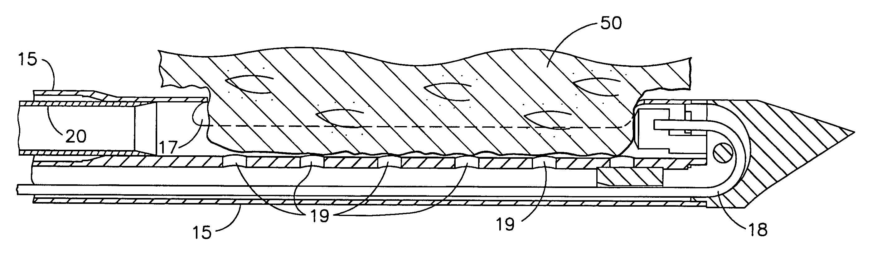
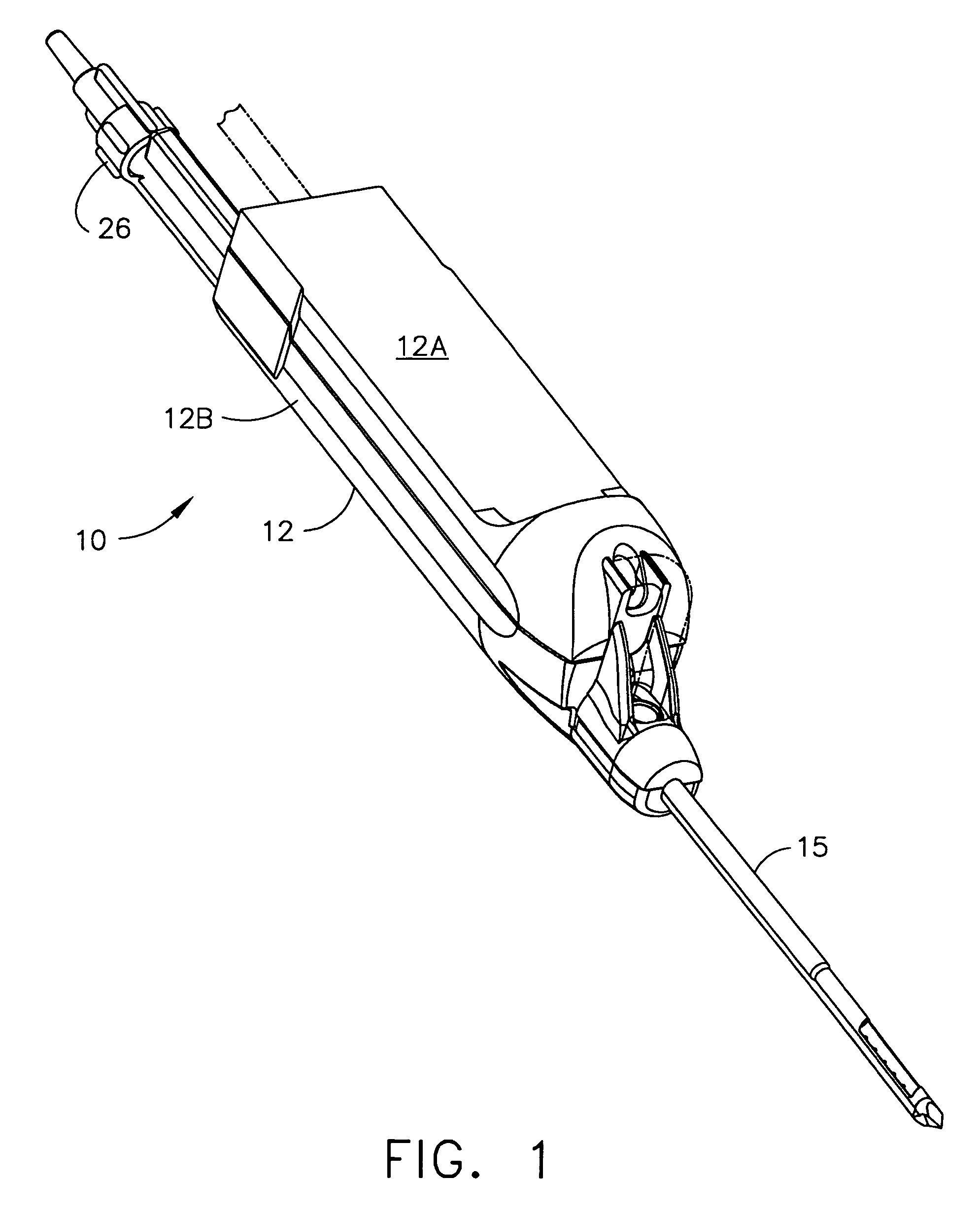
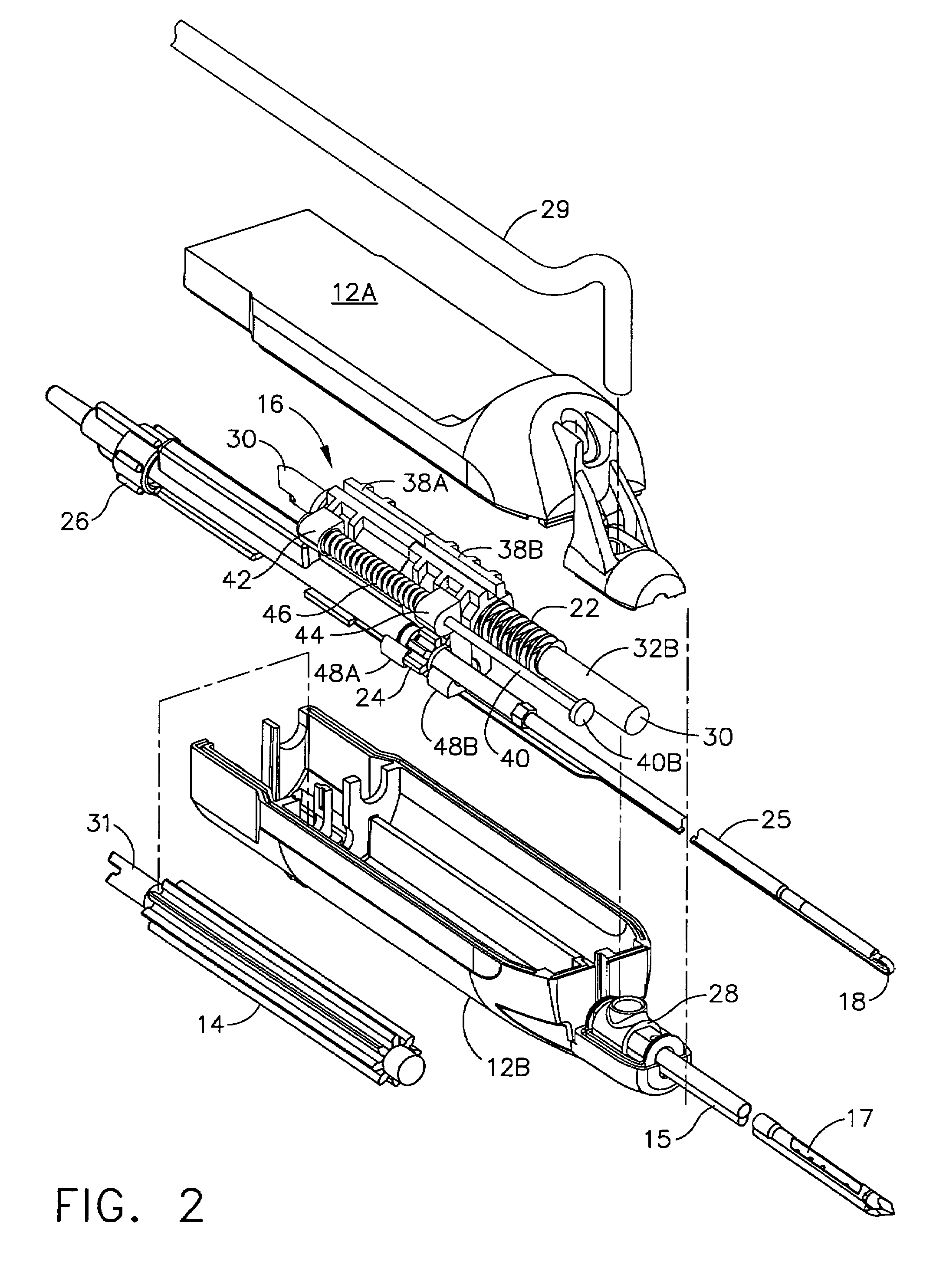
[0061]Referring to FIGS. 1 through 3A, a hand held biopsy instrument 10, embodying the present invention, is illustrated. Biopsy instrument 10 comprises an outer housing 12 comprising a top and bottom shell 12A and 12B respectively. Extending distally outward from bottom shell 12B is biopsy needle 15 the function of which will become apparent below. Contained within housing 12 is drive mechanism 16 for operating the specimen cutter 20 and specimen collector tube 25 subassembly, along with specimen push rod 18 as illustrated in FIG. 3A.

[0062]Specimen collection tube 25 is coaxially positioned within cutter 20 that in turn is coaxially positioned within the upper lumen 13 of the biopsy needle 15 as illustrated in FIGS. 3, 3A, and 4A. Push rod 18 is positioned within the lower lumen 19 within biopsy needle 15 as indicated in FIGS. 3, 3A, and 4A. A vacuum port connector with knockout pin 26, fluidly attached to a vacuum source (not shown), is attached to th...


PUM


Abstract

A biopsy instrument with an internal specimen collection mechanism is provided.

Description

FIELD OF THE INVENTION[0001]The present invention generally relates to instruments for surgically sampling living tissue. More particularly the present invention relates to an improved biopsy probe for acquiring subcutaneous biopsies and / or removing lesions etc.BACKGROUND OF THE INVENTION[0002]The diagnosis and treatment of patients with cancerous tumors, pre-malignant conditions, and other disorders has long been an area of intense investigation. Non-invasive methods for examining tissue include palpation, X-ray, MRI, CT, and ultrasound imaging. When the physician suspects that a tissue may contain cancerous cells, a biopsy may be done using either an open procedure or a percutaneous procedure. For an open procedure, a scalpel is used by the surgeon to create a large incision in the tissue in order to provide direct viewing and access to the tissue mass of interest. The entire mass (excisional biopsy) or a part of the mass (incisional biopsy) may then be removed. For a percutaneous...

Claims


Application Information

Patent Timeline
Patent Type & Authority Patents(United States)
IPC IPC(8): A61B10/00A61B17/14A61B17/32A61B10/02A61B17/34
CPCA61B10/0266A61B10/0275A61B2010/0225A61B2010/0208A61B10/0283A61B10/00
Inventor HIBNER, JOHNPRIVITERA, SALVATOREHUITEMA, THOMAS W.
Owner DEVICOR MEDICAL PROD
Why Eureka
Try Eureka