Supercharge Your Innovation With Domain-Expert AI Agents!

Automated imaging device and method for registration of anatomical structures

an anatomical structure and automatic imaging technology, applied in the field of automatic imaging devices and methods for registration of anatomical structures, can solve problems such as not being the case, and achieve the effect of reducing the exposure of patients to harmful radiation

Active Publication Date: 2015-02-03
BRAINLAB
View PDF8 Cites 6 Cited by
  • Summary
  • Abstract
  • Description
  • Claims
  • Application Information

AI Technical Summary

Benefits of technology

This approach reduces the number of images needed, minimizes patient exposure to radiation, and improves the accuracy of anatomical modeling by optimizing imaging conditions for specific parts, enabling efficient capture of three-dimensional models.

Problems solved by technology

In the first image, however, this is often not the case.

Method used


Image

  • Automated imaging device and method for registration of anatomical structures
  • Automated imaging device and method for registration of anatomical structures
  • Automated imaging device and method for registration of anatomical structures

Examples


Embodiment Construction

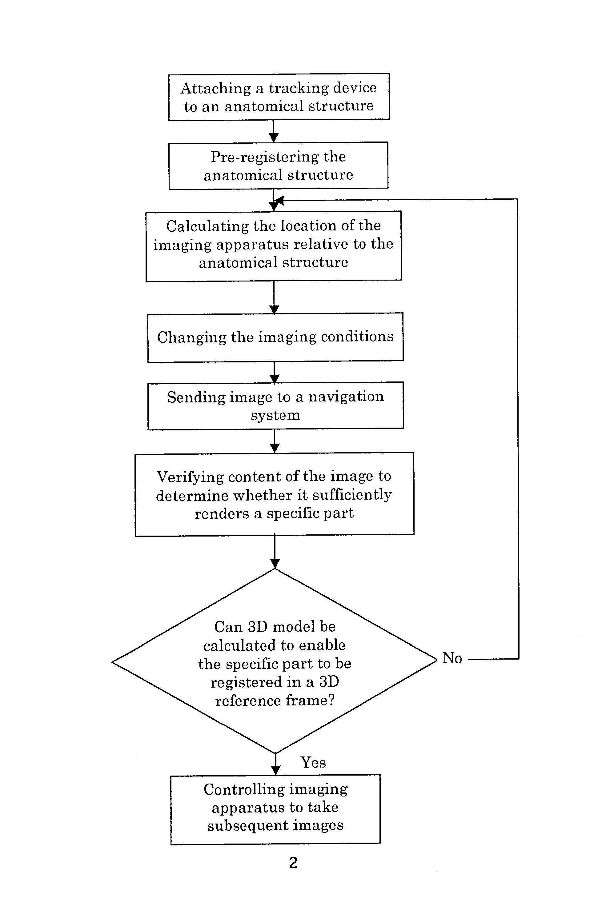
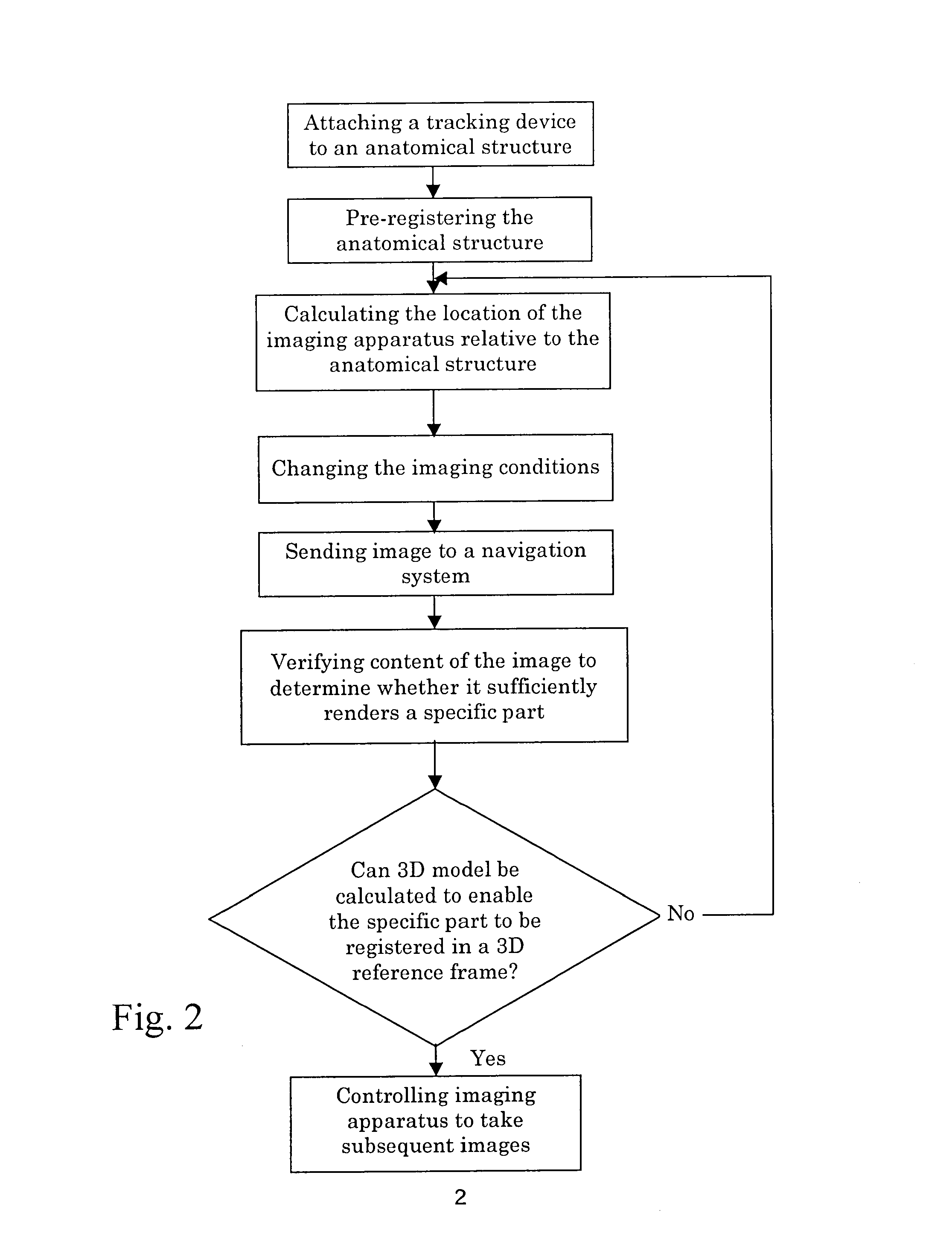
[0041]The method in accordance with the invention can allow a specific part of an anatomical structure, or a three-dimensional model of the specific part, to be captured by an imaging apparatus. The three-dimensional model may be registered using the position of the three-dimensional model in a reference frame determined using a navigation system.

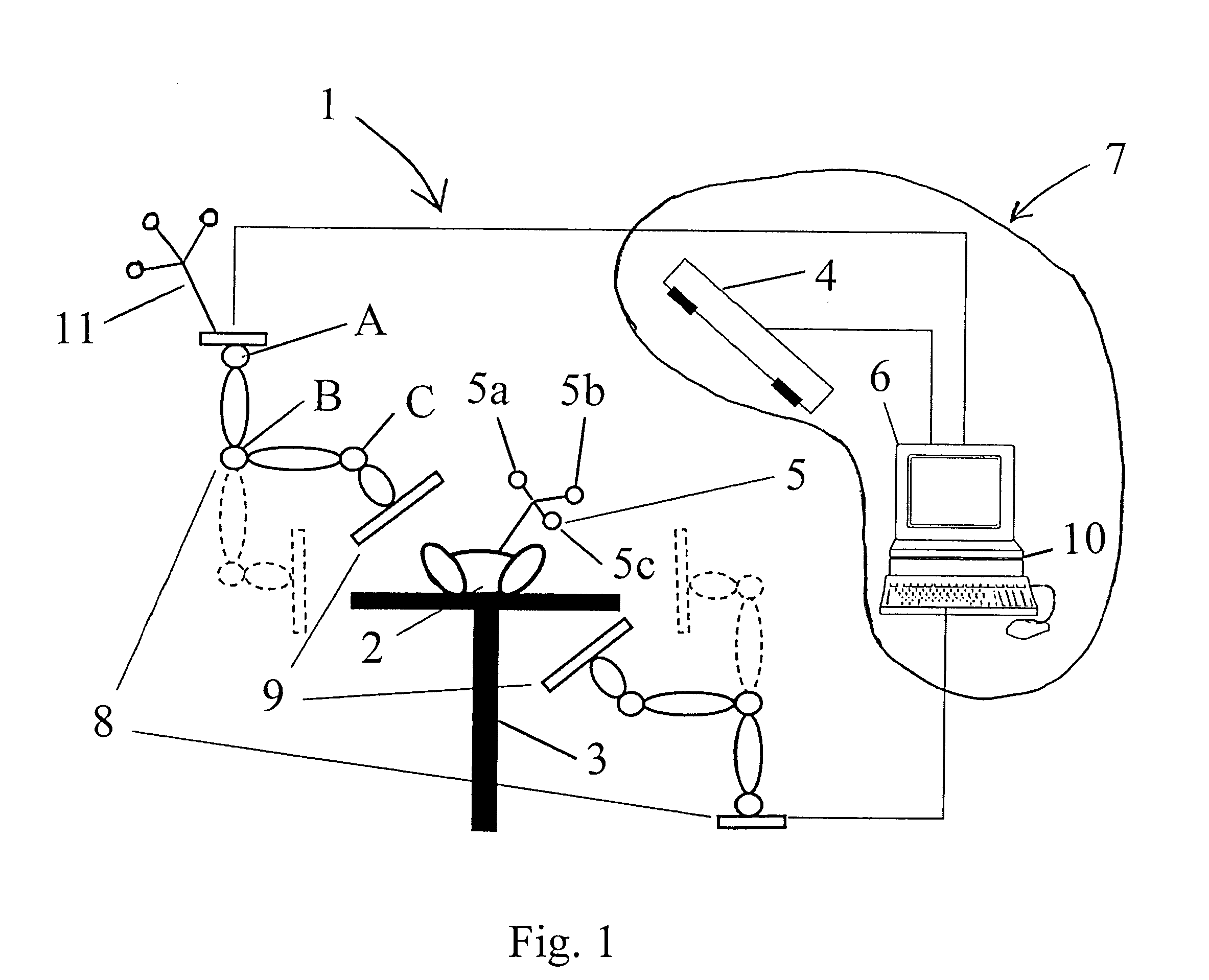
[0042]In addition to one or more of the steps of the method described above in connection with EP 1 611 863, the method in accordance with the invention includes one or more of the following steps[0043]Calculate optimum positions of an imaging apparatus (e.g. an x-ray machine in a C-arm), based on a rough three-dimensional model of the anatomical structure (or the specific part) and navigation data (captured using a sensor and / or a navigation system), wherein the navigation data may be particular data that has been captured using a tracking device and / or pointers that are attached or secured in relation to the anatomical structure.[0044]The...


PUM


Abstract

A device and method for capturing images of a specific part of an anatomical structure using a series of images, wherein an image is taken of the anatomical structure and is compared to a database of information is order to determine and optimize the imaging conditions used for taken subsequent images of the specific part.

Description

RELATED APPLICATION DATA[0001]This application claims priority from Provisional Ser. No. 60 / 887,976, filed Feb. 2, 2007, and EP 07 001 290.1, filed Jan. 22, 2007, which are incorporated by reference.FIELD OF THE INVENTION[0002]The present invention relates to methods and devices for capturing a specific part of an anatomical structure of a human or animal by taking a series of images.BACKGROUND OF THE INVENTION[0003]In past medical treatment practice, when images are taken of a patient, the patient may be lying on a couch and an imaging apparatus such as an x-ray machine may be next to the patient. This x-ray machine may be positioned at a determined location relative to the couch and may be used to image a specific part of the anatomical structure of the patient, for example the pelvis. Often, the specific part is not captured by the first image. The physician then corrects the location of the x-ray machine and takes a second image. If the second image again does not capture the sp...

Claims


Application Information

Patent Timeline
Patent Type & Authority Patents(United States)
IPC IPC(8): G06K9/00A61B6/00A61B19/00
CPCA61B6/488A61B6/4476A61B6/469A61B6/547A61B19/52A61B19/5244A61B19/50A61B2019/501A61B2019/505A61B2019/5255A61B2019/566Y10S128/92Y10S128/922Y10S128/923A61B2034/101A61B90/36A61B2034/2055A61B2034/256A61B34/20A61B34/10A61B2034/105
Inventor WOHLGEMUTH, RICHARDTUMA, GREGOR
Owner BRAINLAB
Features
  • R&D
  • Intellectual Property
  • Life Sciences
  • Materials
  • Tech Scout
  • Unparalleled Data Quality
  • Higher Quality Content
  • 60% Fewer Hallucinations
Learn More