Coronary artery CT (computed tomography) contrastographic image calcification point detecting method

A technique for coronary artery and angiographic images, which is applied in the application field of image processing technology in the medical field, can solve the influence of volume effect, automatically and accurately locate vascular calcification points, differences and other problems, and achieve the effect of overcoming part of the volume effect

Inactive Publication Date: 2013-10-02
SOUTHEAST UNIV
View PDF4 Cites 24 Cited by
  • Summary
  • Abstract
  • Description
  • Claims
  • Application Information

AI Technical Summary

Problems solved by technology

[0003] Since the heart chambers and blood vessels in the coronary CT angiography images all show high CT values, but the range of CT values ​​varies greatly with different parameters such as image acquisition and contrast agent injection, and will also be affected by some The influence of volume effect
Therefore, it is difficult to automatically and accurately locate vascular calcification points in such images with the threshold method used to determine the calcification score in CT plain scan images.

Method used


Image

  • Coronary artery CT (computed tomography) contrastographic image calcification point detecting method
  • Coronary artery CT (computed tomography) contrastographic image calcification point detecting method
  • Coronary artery CT (computed tomography) contrastographic image calcification point detecting method

Examples


Embodiment Construction

[0034] The present invention will be further described below in conjunction with the accompanying drawings.

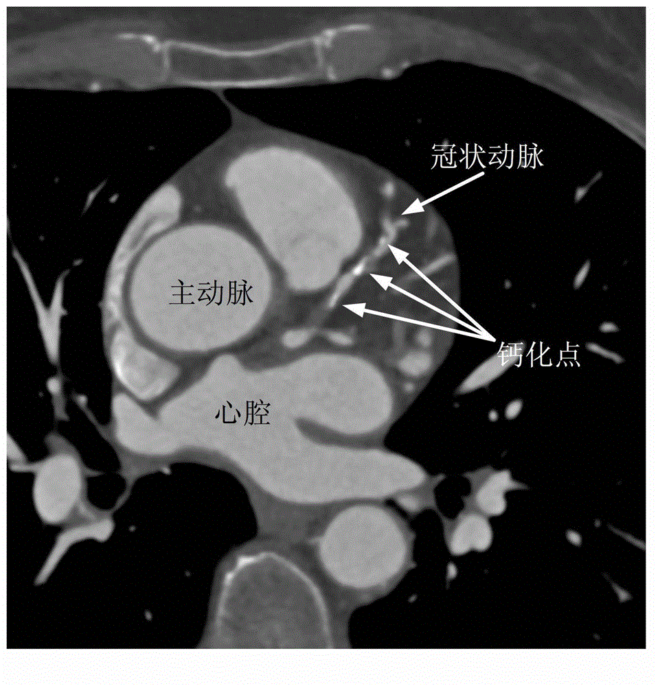
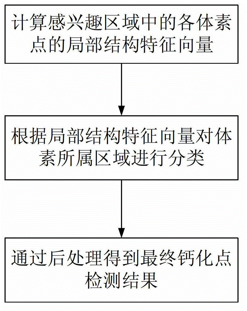
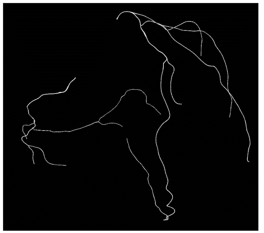
[0035] A method for detecting calcification points in coronary CT angiography images, using the existing central axis of the coronary artery, first extracting the local structural features of each voxel point in the region of interest of the blood vessel, and then using spherical harmonic function transformation to quantify the local structural features The eigenvectors are obtained, and finally the classification algorithm is used to classify the obtained eigenvectors to determine the similarity between the voxel points and the image background, vascular lumen and calcification points in the training data set, and finally obtain the calcification point detection results.

[0036] The steps for generating the region of interest are:

[0037] a. Interpolate the three-dimensional coronary CT angiography image to obtain three-dimensional volume data with equal resolution...


PUM


Abstract

The invention discloses a coronary artery CT (computed tomography) contrastographic image calcification point detecting method. According to the method, the existing coronary artery central axis is utilized, firstly, the local structural features of each voxel point in an interested region of a blood vessel are extracted, then, the spherical harmonic conversion is utilized for quantizing the local structural features, feature vectors are obtained, finally, a classification algorithm is adopted for classifying the obtained feature vectors, the local structural feature approximation degree of the voxel points to the calcification points, blood vessel lomens and image backgrounds in a training dataset is determined, and calcification point detecting results are finally obtained. The method has the advantages that calcification points positioned on the coronary artery blood vessel walls in the coronary artery CT contrastographic image can be precisely positioned, and the efficiency and the accurate rate of the computer auxiliary diagnosis are improved.

Description

technical field [0001] The invention relates to a method for detecting calcification points in coronary CT angiographic images, which belongs to the application of image processing technology in the medical field. Background technique [0002] Coronary heart disease is a serious disease that threatens human health. For many years, coronary heart disease has been ranked among the top causes of human death both at home and abroad. Calcifications located on coronary arteries are an important clinical symptom of coronary heart disease. Correct identification of calcifications and the severity of calcifications will help to accurately diagnose coronary heart disease and formulate a final treatment plan. For this reason, ordinary CT plain scan method is used clinically to measure coronary artery calcium scoring (Calcium scoring) without injection of contrast agent. However, in recent years, with the widespread use of coronary artery CT angiography imaging, doctors can observe the...

Claims


Application Information

Patent Timeline
Patent Type & Authority Applications(China)
IPC IPC(8): G06T17/00A61B6/03
Inventor 杨冠羽王征伍家松杨淳沨舒华忠
Owner SOUTHEAST UNIV
Why Eureka
Try Eureka