
Thyroid CT image computer-aided diagnosis system and method
A computer-aided CT image technology, applied in the field of medical image analysis, can solve the problems of low detection rate of thyroid disease and insufficient standard treatment rate, and achieve the effect of improving identification accuracy, avoiding the influence of human subjective factors, and low cost
- Summary
- Abstract
- Description
- Claims
- Application Information
AI Technical Summary
Problems solved by technology
Method used
Image

Examples
Embodiment
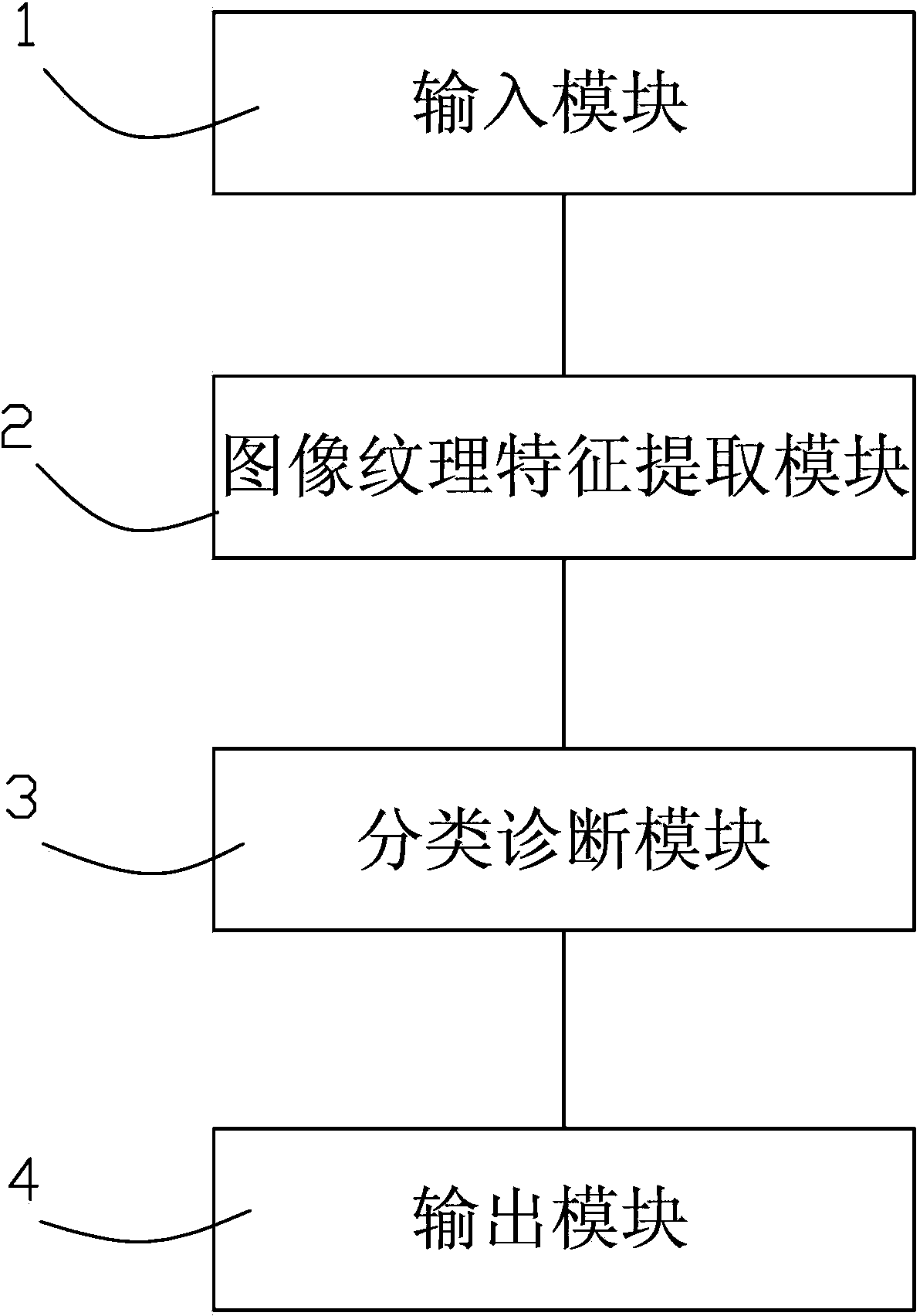
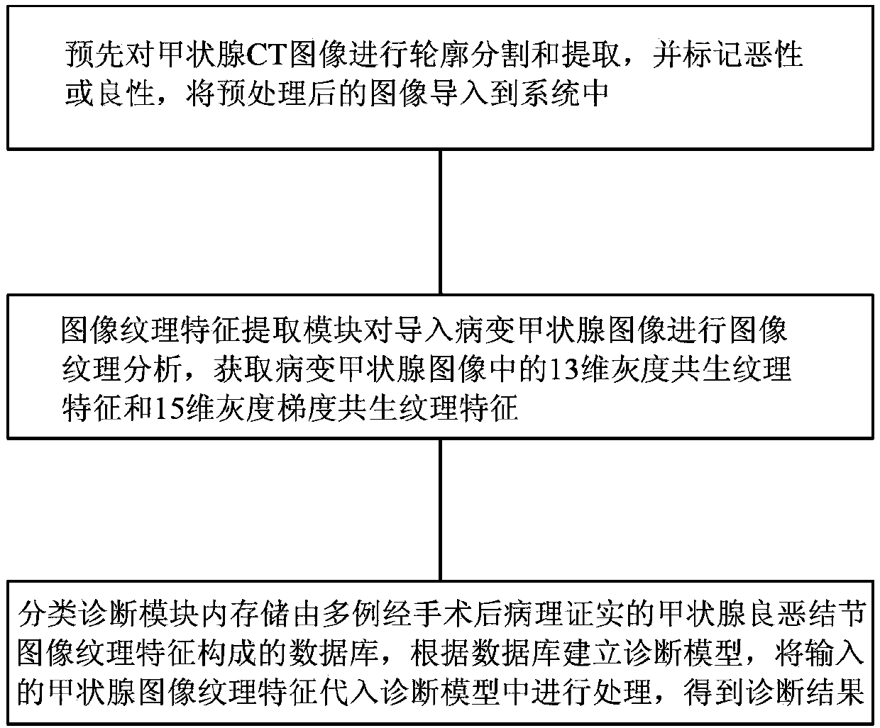
[0110] Present embodiment a kind of thyroid CT image computer-aided diagnosis system, such as figure 1 As shown, it includes an input module 1 , an image texture feature extraction module 2 , a classification diagnosis module 3 and an output module 4 connected in sequence. This input module is used to import thyroid images after contour segmentation and extraction. The image texture feature extraction module performs image texture analysis on the extracted thyroid image containing lesions to obtain the image features of the target image, and the image features are 13-dimensional grayscale co-occurrence texture features and 15-dimensional gray-scale gradient co-occurrence texture features. The classification diagnosis module includes a classifier in the module. The module contains multiple cases of benign and malignant thyroid CT image texture feature databases that have been pathologically diagnosed after surgery. According to the diagnostic model established in the existing d...
PUM
 Abstract
Description
Claims
Application Information