Fluorescence in situ hybridization detection method of IncRNA in esophageal squamous carcinoma tissues

A technique of fluorescence in situ hybridization and esophageal squamous cell carcinoma, which is applied in the field of fluorescence in situ hybridization detection, can solve the problems of poor imaging quality of lncRNA hybridization color development, etc., and achieve the effect of low cost, simple operation and avoiding interference

Inactive Publication Date: 2017-01-25
ZHONGSHAN HOSPITAL FUDAN UNIV
View PDF8 Cites 5 Cited by
  • Summary
  • Abstract
  • Description
  • Claims
  • Application Information

AI Technical Summary

Problems solved by technology

However, non-specific color development of DAB staining in tumor tissue is ...

Method used


Image

  • Fluorescence in situ hybridization detection method of IncRNA in esophageal squamous carcinoma tissues
  • Fluorescence in situ hybridization detection method of IncRNA in esophageal squamous carcinoma tissues

Examples


Embodiment 1

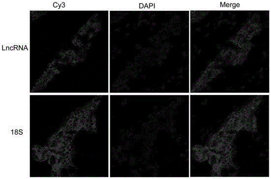
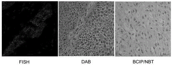
[0022] Example 1: Fluorescence in situ hybridization detection method of lncRNA in esophageal squamous cell carcinoma tissue

[0023] 1. Reagent preparation:

[0024] 1) lncRNA FISH Probe Mix storage solution (20 μM), return to room temperature (RiboBio, Cat. No. C10920), the probe is labeled with cy3.

[0025] 2) U6, 18s internal reference FISH Probe Mix stock solution (20 μM), return to room temperature (RiboBio, product number lnc110101, lnc110102).

[0026] 3) Pre-hybridization solution: mix an appropriate amount of Blocking Solution (Ribobio, product number: C10903) and 1×Pre-hybridization Buffer (Ribobio, product number: C10901) at a ratio of 1:99, and incubate at 37°C until transparent After clarifying the state, aliquot and save. Incubate in an air or water bath at 37°C until clear and transparent before use.

[0027] Note: 1×Pre-hybridization Buffer may precipitate when it is taken out from the low temperature, which is a normal phenomenon.

[0028] 4) Hybridizati...


PUM


Abstract

The invention provides a fluorescence in situ hybridization detection method of IncRNA in esophageal squamous carcinoma tissues. The method is characterized by comprising the following steps of preparing frozen esophageal squamous carcinoma tissue slices; performing fixation, digestion and penetration; using IncRNA probes for detection; performing DNA dyeing and chip sealing; using confocal microscopy detection imaging or fluorescence microscope detection imaging. The fluorescence in situ hybridization detection method of IncRNA in the esophageal squamous carcinoma tissues provided by the invention has the advantages that the cost is low; the operation is simple; the time and the labor are saved. The method can be used for conveniently detecting the expression level of the IncRNA in the esophageal squamous carcinoma tissues; the positioning conditions of the IncRNA in sub cells in the esophageal squamous carcinoma tissues can be directly shown.

Description

technical field [0001] The invention relates to a fluorescence in situ hybridization detection method for lncRNA in esophageal squamous cell carcinoma tissue. Background technique [0002] The in situ detection technology of lncRNA in esophageal squamous cell carcinoma cell lines is relatively mature, but the fluorescence in situ hybridization technology of lncRNA in esophageal squamous cell carcinoma tissue has not been mature. Due to the restriction of collagen and cellulose in the stroma of esophageal squamous cell carcinoma, too much fluorescent agent remains, the background staining is poor, and the distinction between tumor parenchyma staining and stroma residue is not obvious. Therefore, the fluorescence in situ hybridization detection technology of lncRNA in esophageal squamous cell carcinoma tissue needs to be further optimized. [0003] Quantitative analysis of lncRNA in esophageal squamous cell carcinoma tissue is commonly used by RT-PCR; the common method of sub...

Claims


Application Information

Patent Timeline
IPC IPC(8): C12Q1/68
CPCC12Q1/6841C12Q2545/101C12Q2563/107C12Q2565/518
Inventor 王帅时雨刘红
Owner ZHONGSHAN HOSPITAL FUDAN UNIV
Why Eureka
Try Eureka