Head and neck angiography method

An angiography, head and neck technology, applied in the direction of cardiac catheterization, medical science, radiodiagnostic equipment control, etc., can solve the problems of increased cost of angiography, large increase in radiation dose, inaccurate control of the amount of contrast agent and realization of individualization, etc., to achieve The operation process is efficient and simple, the strengthening effect is good, and the effect of reducing the incidence of allergic reactions and radiation damage

Pending Publication Date: 2020-10-20
CHONGQING TRADITIONAL CHINESE MEDICINE HOSPITAL
View PDF0 Cites 2 Cited by
  • Summary
  • Abstract
  • Description
  • Claims
  • Application Information

AI Technical Summary

Problems solved by technology

Disadvantages: Although the final image obtained by this inspection method can meet the diagnostic requirements, the radiation dose increases greatly, and the amount of contrast agent cannot be accurately controlled and personalized.
Therefor

Method used


Image

  • Head and neck angiography method
  • Head and neck angiography method
  • Head and neck angiography method

Examples


Embodiment 1

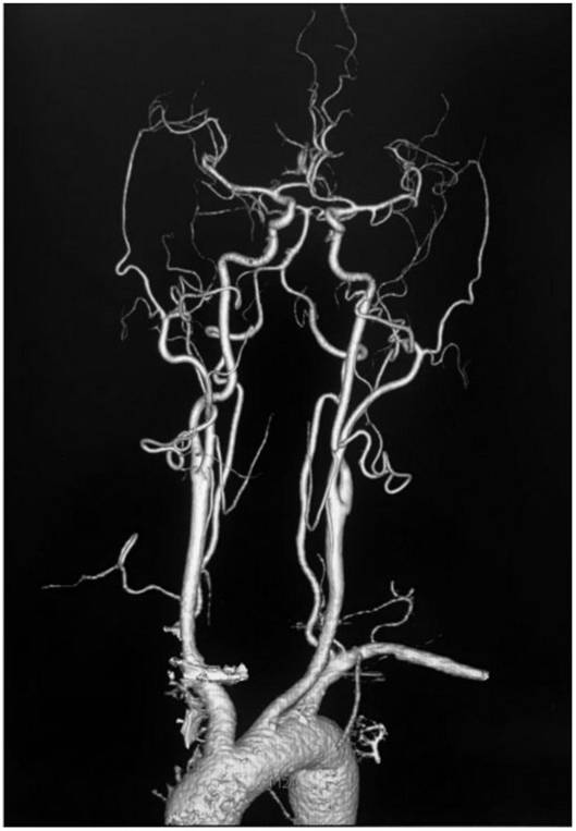
[0046] Refer to attached figure 1 , 1) Obtain basic data: obtain the basic data of subject 1, the basic data include gender: male, set male=1, age 65, heart rate 79 beats / min, systolic blood pressure 128mmHg and diastolic blood pressure 67mmHg;

[0047] 2) Calculation of peak time PT: Iterate the data of the subject obtained in step 1) into the PT time calculation formula: PT=22.860+0.055*65+1.591*1-0.080*79-0.029*128+0.041*67 ; The calculated PT time is 20.741 seconds, and the PT time with one decimal place is 20.7 seconds.

[0048] 3) Determining the scanning / exposure time T: according to the specific inspection purpose of the examinee 1, the scanning range is defined as head and neck blood vessels, and the scanning / exposure time is determined to be 5.1 seconds through the defined scanning range;

[0049] 4) Calculate the total amount of contrast agent: According to step 2), the peak time PT calculated in step 2) is 20.7 seconds, and the scanning / exposure time determined in...

Embodiment 2

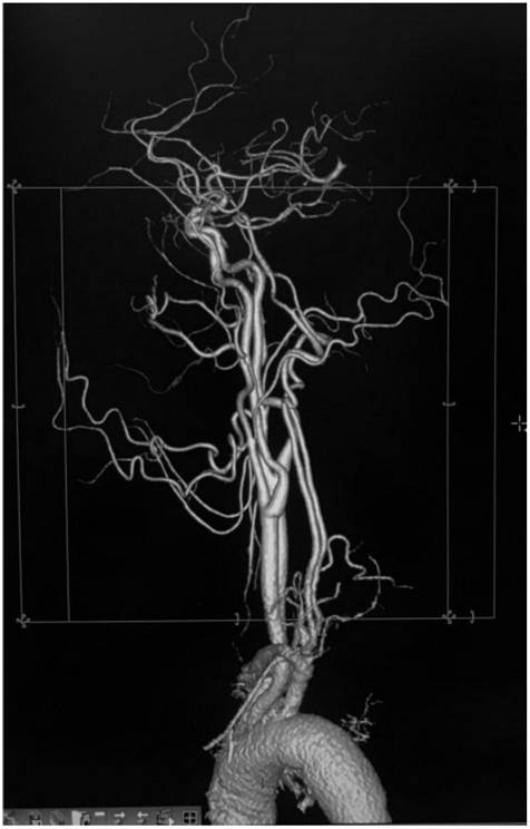
[0053] Refer to attached figure 1 , 1) Obtain basic data: obtain the basic data of the person to be examined 2, the basic data include gender: male, set male=1, age 60 years old, heart rate 78 beats / min, systolic blood pressure 135mmHg and diastolic blood pressure 78mmHg;

[0054] 2) Calculate the peak time PT time: iterate the data of the subject obtained in step 1) into the PT time calculation formula: PT=22.860+0.055*60+1.591*1-0.080*78-0.029*135+0.041* 78; The calculated PT time is 20.794 seconds, and the PT time with one decimal place is 20.8 seconds.

[0055] 3) Determine the scanning / exposure time T: according to the specific inspection purpose of the examinee 2, the scanning range is defined as the head and neck blood vessels, and the scanning / exposure time is determined to be 5.1 seconds through the defined scanning range;

[0056] 4) Calculate the total amount of contrast agent: According to the calculation in step 3), the peak time PT is 20.8 seconds, and the scann...

Embodiment 3

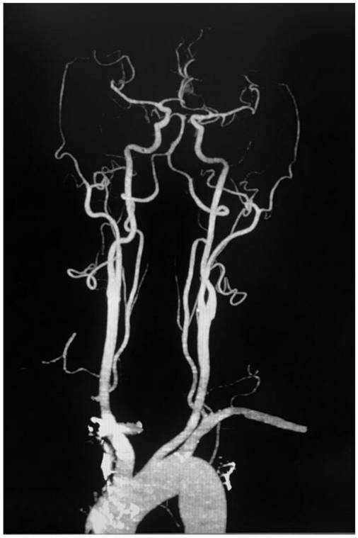
[0060] Refer to attached figure 1 , 1) Obtain basic data: obtain the basic data of the person to be examined 3, the basic data include gender: male, set male=1, age 77 years old, heart rate 79 beats / min, systolic blood pressure 138mmHg and diastolic blood pressure 90mmHg;

[0061] 2) Calculate peak time PT: Iterate the data of the subject obtained in step 1) into the PT time calculation formula: PT=22.860+0.055*77+1.591*1-0.080*79-0.029*138+0.041*90 ; The calculated PT time is 22.054 seconds, and the PT time with one decimal place is 22.1 seconds.

[0062] 3) Determine the scanning / exposure time T: according to the specific inspection purpose of the examinee 3, the scanning range is defined as the head and neck blood vessels, and the scanning / exposure time is determined to be 5.1 seconds through the defined scanning range;

[0063] 4) Calculate the total amount of contrast agent: According to the calculation in step 2), the peak time PT is 22.1 seconds, and the scanning / expos...


PUM


Abstract

The invention relates to the field of medical imaging, and in particular relates to a head and neck angiography method. The head and neck angiography method comprises the following steps of: (1), obtaining the basic data of an examined person including the sex, the age, the heart rate, the systolic pressure and the diastolic pressure; (2), substituting the obtained basic data of the examined person into a PT time calculation formula; (3), determining the scanning/exposure time T through a defined scanning range; (4), obtaining the total injection time according to the PT time and the exposuretime T, selecting the injection rate of a contrast agent to be 3.0-5.0 ml/s, and obtaining the total injection amount and the total amount of the contrast agent by calculation; and (5), setting the delay time to the PT time on a CT scanner, starting the exposure key of the CT scanner and a contrast agent injector at the same time, injecting the total amount of the contrast agent, then, continuously injecting the total amount of normal saline injection, and performing exposure by the CT scanner, so that an examination image is obtained. The head and neck angiography method in the invention is used for improving the imaging effect of blood vessels; the use amount of the contrast agent is used in a personalized manner; therefore, the radiation dosage is reduced; and the radiation injury of radiography on a patient and the rate of allergic reaction are reduced.

Description

technical field [0001] The invention relates to the field of angiography methods of medical imaging technology, in particular to an angiography method used in the examination of blood vessels in the head and neck. Background technique [0002] Cerebrovascular disease is a common neurological disease in clinical practice. In recent years, its incidence has gradually increased and its mortality rate is high. However, 50%-70% of the survivors of cerebrovascular disease have sequelae, which increases the burden on society and family. In the existing technology, combined head and neck CT angiography (Computed Tomography Angiography, CTA) can clearly display the vascular images of the common carotid artery, internal and external carotid artery, vertebral artery, basilar artery and the circle of Willis, and can distinguish the relevant Therefore, CTA has been widely used in the diagnosis of head and neck vascular lesions. In the process of CTA angiography, in order to make head an...

Claims


Application Information

Patent Timeline
IPC IPC(8): A61B6/03A61B6/00
CPCA61B6/032A61B6/481A61B6/504A61B6/54A61B6/542
Inventor 张德川杨华王贵学范龙玲冯晨刘曦
Owner CHONGQING TRADITIONAL CHINESE MEDICINE HOSPITAL
Why Eureka
Try Eureka