Intraoperative pulmonary nodule positioning device in pleural cavity and preparation method thereof

A technology for positioning devices and pulmonary nodules, which is applied in the field of medical devices, can solve the problems of increasing radiation dose, increasing the economic burden of patients, and high cost of imaging department puncture management, achieving the effect of economical positioning methods and reducing economic burden

Active Publication Date: 2022-06-24
SHANGHAI PULMONARY HOSPITAL
View PDF19 Cites 0 Cited by
  • Summary
  • Abstract
  • Description
  • Claims
  • Application Information

AI Technical Summary

Problems solved by technology

[0003] In order to accurately remove pulmonary nodules under thoracoscopic surgery, percutaneous Hook-wire positioning, percutaneous coil positioning and liquid dye injection positioning under the guidance of traditional preoperative CT are gradually popularized, but in clinical practice It was found that this positioning process has many disadvantages: (1) The patient needs to undergo multiple CT scans, which increases the radiation dose and increases the economic burden of the patient; (2) There may be unavoidable pneumothorax, blood Chest, pleural reaction and iatrogenic injury risk of thoracic great vessels; (3) The traditional positioning mode requires high coordination and cooperation in imaging departments, operating rooms, etc. In general hospitals, the cost of puncture investment and management in the imaging department is relatively high

Method used


Image

  • Intraoperative pulmonary nodule positioning device in pleural cavity and preparation method thereof
  • Intraoperative pulmonary nodule positioning device in pleural cavity and preparation method thereof

Examples


Embodiment

[0029] The present embodiment provides a method for preparing an intraoperative pulmonary nodule positioning device in the pleural cavity, the steps comprising:

[0030] S1. Based on the CT image data of the patient's chest, construct a three-dimensional digital model of the patient's lungs and thorax;

[0031] S2. Based on the three-dimensional digital model, prepare the intraoperative pulmonary nodule positioning device in the pleural cavity;

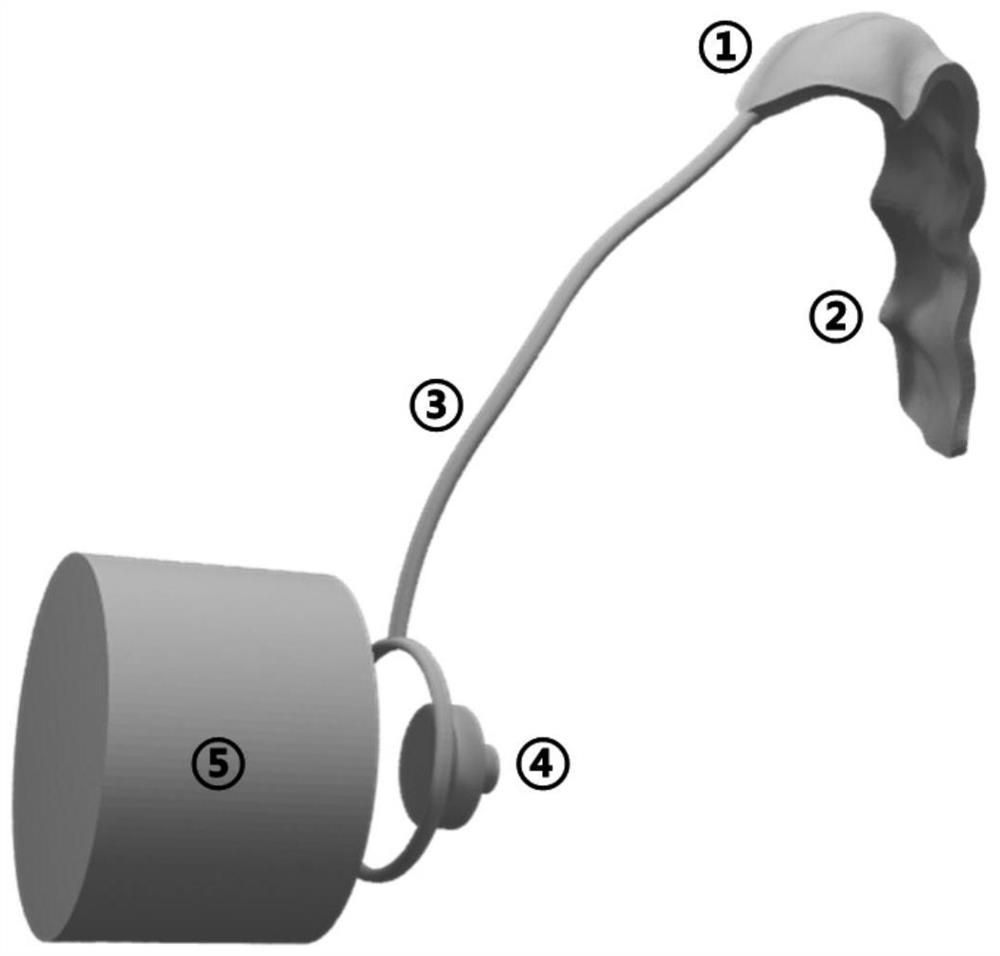
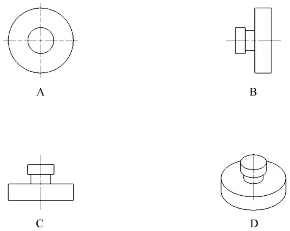
[0032] Wherein, the intraoperative pulmonary nodule positioning device in the pleural cavity includes: a fixing assembly 2, a positioning assembly 1 and an arcuate positioning arm 3 connected in sequence; the positioning assembly 1 is matched with the apex of the patient's thoracic cavity; the fixing assembly 2 is matched with the upper thoracic vertebra of the patient; the arcuate positioning arm 3 is matched with the patient's thorax, and the end of the arcuate positioning arm 3 is a ring structure, and the center of the ring struct...


PUM


Abstract

The present invention relates to a preparation method of a device for locating pulmonary nodules in the pleural cavity during operation. The intraoperative pulmonary nodule positioning device in the pleural cavity; wherein, the intraoperative pulmonary nodule positioning device in the pleural cavity includes: a fixed assembly, a positioning assembly, and a bow-shaped positioning arm connected in sequence; the positioning assembly is connected to the patient's chest lung apex The fixed assembly matches the patient's upper thoracic vertebrae; the arc positioning arm matches the patient's chest, and the end of the arc positioning arm is a ring structure, and the center of the ring structure is aligned with the positioning point on the patient's lung surface match. The invention provides a safer, more accurate, more convenient and economical positioning method for patients and clinicians.

Description

technical field [0001] The invention relates to the technical field of medical devices, in particular to an intraoperative pulmonary nodule positioning device in a pleural cavity and a preparation method thereof. Background technique [0002] Lung cancer is one of the malignant tumors with the highest morbidity and mortality in my country. With the popularization of low-dose computed tomography (LDCT) lung cancer screening, the sensitivity of early-stage lung cancer diagnosis has improved, and more and more solitary lung cancers have been diagnosed. Small nodules (<1 cm in diameter) were found. Because such small nodules have a certain malignant probability, their diagnosis and treatment are the focus of clinicians. Video-assisted thoracoscopic surgery (VATS) provides a minimally invasive way to diagnose and treat small pulmonary nodules, but endoscopic operators have limited visual field and operating space, and it is often difficult to quickly locate the nodule during s...

Claims


Application Information

Patent Timeline
Patent Type & Authority Patents(China)
IPC IPC(8): A61B17/34A61B90/00
CPCA61B17/3403A61B90/39
Inventor 陈昶汤海孙维言佘云浪王龙范子文鄂浩然陈家飞
Owner SHANGHAI PULMONARY HOSPITAL
Why Eureka
Try Eureka