

Medical visual puncture needle
A puncture needle and needle core technology, applied in the direction of puncture needle, application, trocar, etc., can solve the problems of misplaced into other tissues and organs, inaccurate puncture position, damage to blood vessels, etc., and achieves convenient operation, low cost of use, and design reasonable effect
Pending Publication Date: 2022-03-11
BEIJING NEUROSURGICAL INST
View PDF0 Cites 0 Cited by
- Summary
- Abstract
- Description
- Claims
- Application Information
AI Technical Summary
Problems solved by technology
However, there are often no auxiliary positioning methods such as imaging and ultrasound in clinical puncture operations. The positioning mainly depends on the experience of clinicians. Therefore, there may be inaccurate puncture positions, the possibility of mistakenly entering other tissues and organs, and the possibility of bleeding caused by damage to blood vessels.
Method used
View moreImage
Smart ImageViewing Examples

Examples
Embodiment Construction
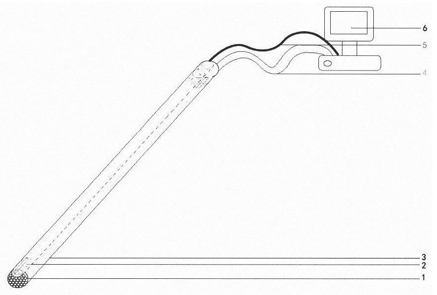
[0007] A new type of visual puncture needle. The needle core of the puncture needle contains an optical fiber, which illuminates and records the puncture path, and makes the path clear. During the puncture operation, the puncture needle is connected to the display screen, so that the picture of the tip of the puncture needle is transmitted to the display screen in real time.
Login to View More PUM
 Abstract
The invention relates to a medical visual puncture needle. The puncture needle core contains light-guide fibers, and a small lamp bead is installed at the tip end of the puncture needle core for illumination. During puncture operation, the puncture needle is connected to the display screen, and pictures of the tip end of the puncture needle are transmitted to the display screen in real time. The in-vivo tissue structure can be found easily, and related negative effects caused by inaccurate puncture positions can be avoided to a great extent. The visual puncture needle has the obvious beneficial effects that firstly, the design is reasonable, related negative effects caused by puncture can be reduced to a great extent under a visual condition, the in-vivo structure can be found, the medical experience of a patient can be effectively improved, and the vital interests of the patient can be guaranteed; operation is convenient, and the use mode is the same as that of a traditional puncture needle. And thirdly, the use cost is low, and repeated use can be realized after disinfection.
Description
technical field [0001] The invention belongs to tangible products, specifically a medical visualized puncture needle, which can be used in clinical medical operations to make the puncture path visible, so as to find pathological changes or tissue structures in the body, reduce inaccurate puncture positions, or reduce tissue damage, reduce resulting complications. Background technique [0002] Puncture operation is an important clinical examination method, which is of great significance for disease diagnosis and treatment. Clinically commonly used puncture needles are often composed of a needle core and a sleeve. When puncturing, the needle core is put into the sleeve, and the needle core is pulled out after puncturing to a predetermined position, and then the clinical operation is performed. However, there are often no auxiliary positioning methods such as imaging and ultrasound in clinical puncture operations. The positioning mainly depends on the work experience of clinic...
Claims
Login to View More Application Information
Patent Timeline
 Patent Type & AuthorityApplications(China)
IPC IPC(8): A61B17/34A61B17/94A61B90/30A61B90/00
CPCA61B17/34A61B90/30A61B90/361A61B2090/306A61B2090/3614
Inventor孟凡刚韩春雷王晗王乔刘焕光张建国
OwnerBEIJING NEUROSURGICAL INST