White spot syndrome virus on-spot detection test paper, its preparation and method of application

An on-site detection and virus technology, applied in the intersection of immunology and virology, can solve the problems of inability to achieve rapid, on-site detection, time-consuming, and high sensitivity easily lead to false positives.

Active Publication Date: 2006-12-27
OCEAN UNIV OF CHINA
View PDF0 Cites 0 Cited by
  • Summary
  • Abstract
  • Description
  • Claims
  • Application Information

AI Technical Summary

Problems solved by technology

These methods, the operation procedures are relatively complicated, require certain laboratory equipment, and take a long time. The whole process takes several or even ten hours, which cannot achieve the purpose of rapid and on-site detection.
PCR method has high sensitivity and can be used for the detection of asymptomatic diseased shrimp, but its high sensitivity can easily lead to false positive results; ELISA and dot immunoblotting are currently commonly used detection methods, but the internal Source enzymes can interfere with its detection results, resulting in false positives

Method used


Image

  • White spot syndrome virus on-spot detection test paper, its preparation and method of application
  • White spot syndrome virus on-spot detection test paper, its preparation and method of application

Examples


Embodiment 1

[0018] Described monoclonal antibody E colloidal gold labeling method is:

[0019] (1) Preparation of colloidal gold: preparation of 18-20nm colloidal gold particles, mixing 100ml of 0.01% chloroauric acid solution with 2.5ml of 0.1% sodium citrate, heating to 100°C to make a colloidal gold solution, the pH value of the solution: Adjust to 8.0-8.4 with 0.2% potassium carbonate, set aside;

[0020] (2) Preparation of gold-labeled monoclonal antibody E:

[0021] Add 1 μl of antibody to 1ml of colloidal gold, mix well, add 100 μl of 10% sodium chloride, observe the color change, if it turns blue, it means that the antibody is insufficient, and then continue to increase the amount of antibody until the color of the colloidal gold does not change. On this basis, increase the amount of this antibody by 50-100%, which is the optimal amount of monoclonal antibody labeled with 1ml of colloidal gold. Colloidal gold is labeled with this appropriate amount of monoclonal antibody, and th...

Embodiment 2

[0023] Preparation of glass fiber layer blocks loaded with gold-labeled monoclonal antibody E: Spray the prepared gold-labeled monoclonal antibody E liquid on the glass fiber membrane until the liquid starts to ooze out, freeze-dry, and store at 4°C for use.

Embodiment 3

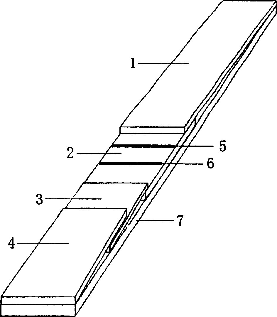
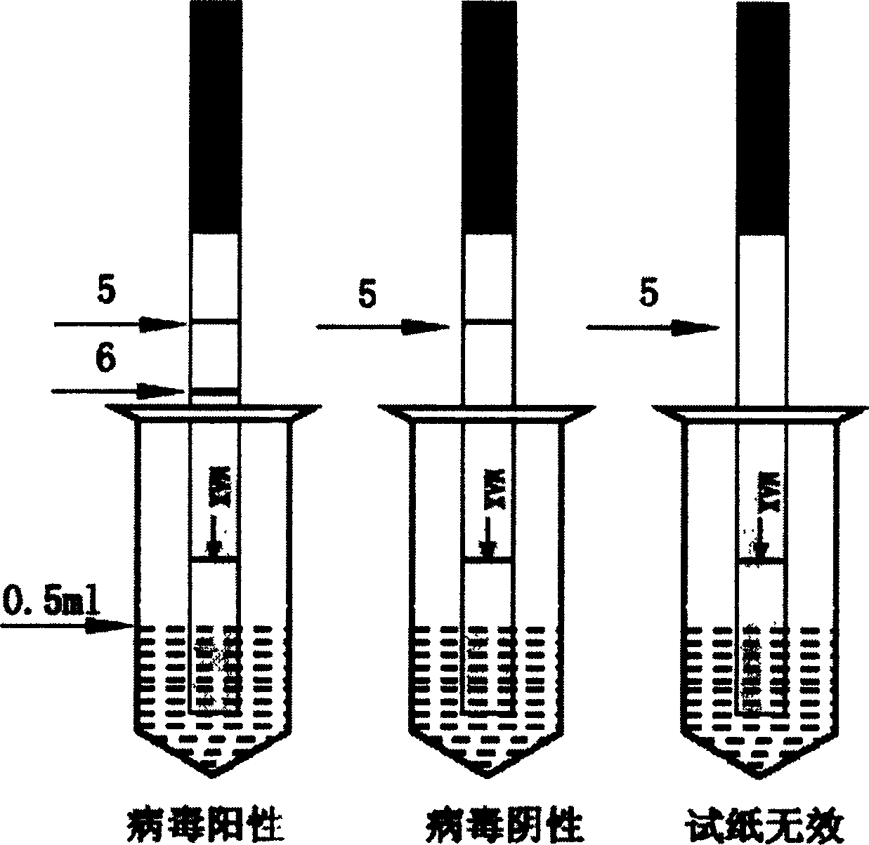
[0025] The preparation method of the detection layer of the white spot virus on-site detection test paper of the present invention is: after the anti-white spot virus monoclonal antibody F purified by the caprylic acid method is freeze-dried, it is prepared into a 4 mg / ml anti-white spot virus monoclonal antibody F liquid, and it is sprayed with a film spraying machine. Spray on the nitrocellulose membrane to form the test line 6; similarly, prepare goat anti-mouse IgG at 300 μg / ml, and spray it on the nitrocellulose membrane with a film spraying machine to form the quality control line 5. The distance between the two lines is 5mm, the quality control line 5 is close to the water-absorbing layer of the hand-held end, and the detection line 6 is close to the water-absorbing layer of the sample loading end. Dry at room temperature, then block with pH 7.4, 0.01M PBS containing 10% bovine serum albumin at 37°C for 30min, rinse with PBS and dry in the air.


PUM


Abstract

The invention relates to an indicator paper for measuring leucodermia virus on line, which comprises: adhesive carrier board; the single antibody E which is colloidal gold marked and secreted by the hybridomas cell whose conserving number is: CCTCC-C200421; the single antibody F and sheep-resistant-mouse IgG which is secreted by the hybridomas cell whose conserving number is: CCTCC-C200423. The ends of the carrier board are fitted with the adding and holding water-absorbing layer, and the middle of the board is fitted with the layer for measuring the nitric fiber film, and in the junction of the layer and the adding water-absorbing layer is fitted with the fiber glass block carrying the E, and one part of the block is set below the water-absorbing layer and the other part is set above the measuring layer, wherein the layer continuing with the block is fitted with the measuring line and quality-controlling line, which are surrounded by the F and IgG..

Description

technical field [0001] The invention relates to the improvement of the detection technology of breeding disease pathogens, specifically a white spot syndrome virus (white spot syndrome virus, WSSV) on-site detection test paper and its preparation and use methods, which belong to the interdisciplinary technical field of immunology and virology. Background technique [0002] White spot virus disease is one of the serious diseases in prawn farming, which leads to high mortality of prawns and huge losses to the prawn farming industry. Given that there is no effective treatment for leukoplakia, rapid and accurate virus isolation and detection technology has become one of the focuses of research by scholars from all over the world. At present, the diagnosis of leukoplakia virus disease mainly depends on the detection of leukoplakia virus in the laboratory. The current laboratory detection methods include: PCR method; DNA probe in situ hybridization method; transmission electron mi...

Claims


Application Information

Patent Timeline
Patent Type & Authority Patents(China)
IPC IPC(8): G01N33/569G01N33/558G01N33/52G01N33/532
Inventor 战文斌王晓洁林颖博
Owner OCEAN UNIV OF CHINA
Why Eureka
Try Eureka