Methods and devices for quantitative analysis of x-ray images

A technology of light image and calibration model, applied in the field of analysis and analysis technology, device of calibration model

Inactive Publication Date: 2004-05-19
IMAGING THERAPEUTICS
View PDF5 Cites 3 Cited by
  • Summary
  • Abstract
  • Description
  • Claims
  • Application Information

AI Technical Summary

Problems solved by technology

However, existing calibration models are not always accurate, either because of the structure or material or because of the calibration model, and also because one or more regions of the calibration model do not always appear on the x-ray image

Method used


Image

  • Methods and devices for quantitative analysis of x-ray images
  • Methods and devices for quantitative analysis of x-ray images
  • Methods and devices for quantitative analysis of x-ray images

Examples


example 1

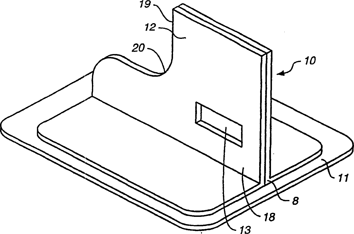
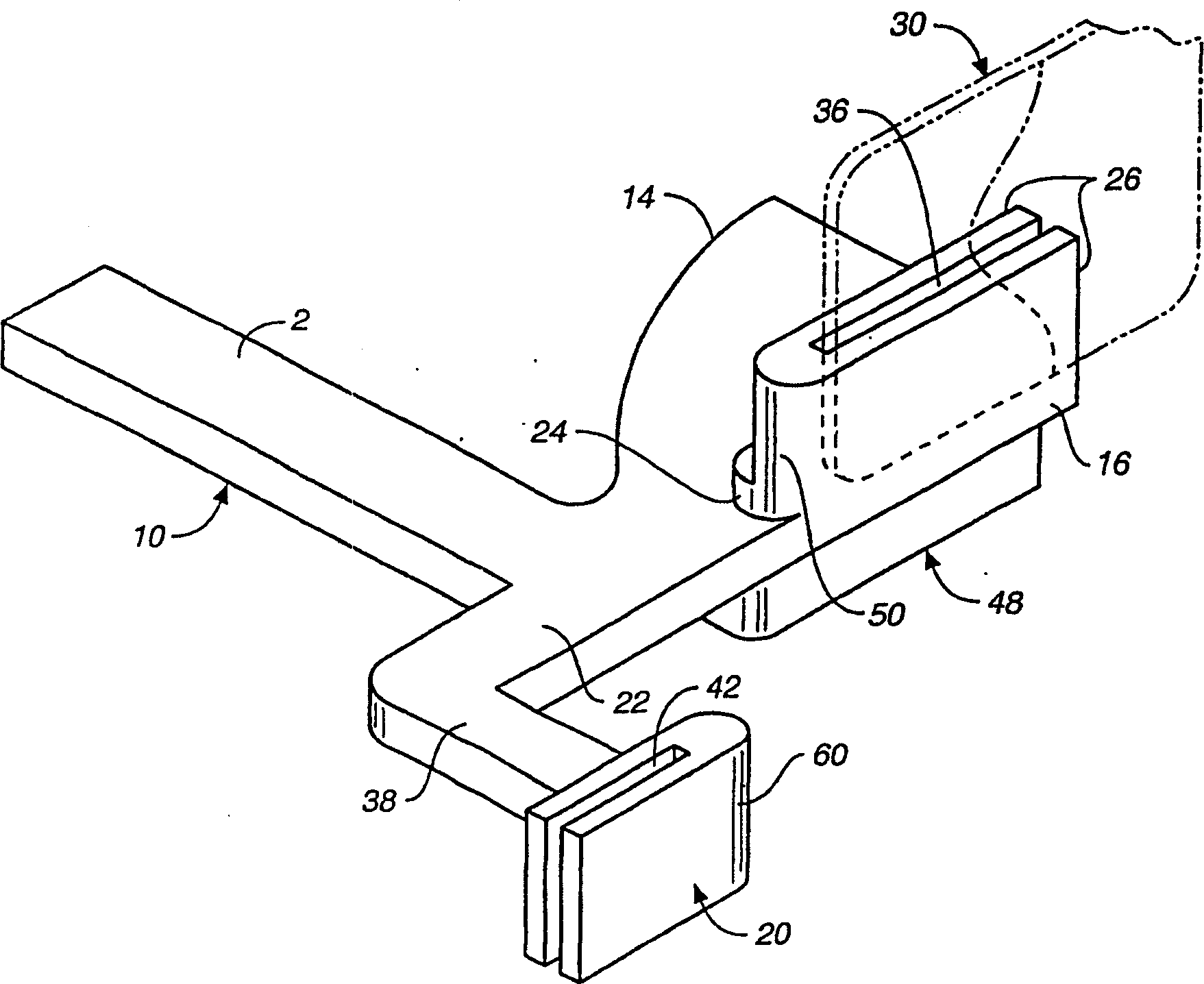
[0133] Example 1: Calibration phantom incorporated into film cover

[0134] The workflow presented here constitutes an example of the use of a calibration model in image acquisition. Professionals can readily recognize other methods of including a calibration model in the acquisition process in order to normalize, standardize, the metrics of the x-ray image.

[0135] In this example, the calibration phantom is incorporated into the dental X-ray film cover that protects the film from light. The film is placed in a film holder containing the film placed in the patient's mouth. The position of the film-filled clip in the patient's mouth is designed in such a way that it ensures that the calibration phantom is not blocked by any structures such as teeth or lips from the X-ray rays.

[0136] After the image was acquired, the film was taken into the darkroom and the cover with the calibration phantom removed. The film is then developed just like regular dental X-ray film.

example 2

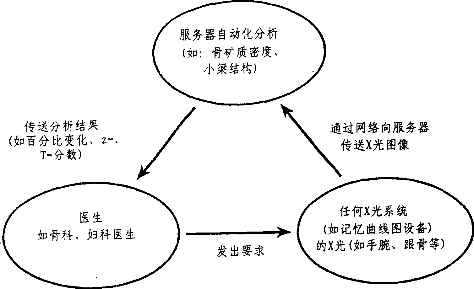
[0137] Example 2: Transmission of X-ray images including calibration models over the network

[0138] This example presents a typical and possible use of the invention; digitized X-images containing calibration phantom images are transmitted over a network. The same use of the present invention can be clearly identified.

[0139] X-ray images are obtained by transmitting the calibration phantom on film. Films with x-ray images and calibration and calibration phantom images are developed. Subsequently, the film is digitized, eg, with a flatbed or slide scanner, resulting in a digital image. The digital images, including the x-ray images and calibration phantom images, are transmitted over the network to a remote computer. The remote computer uses information in the x-ray images and / or calibration phantom images to perform one or more measurements.

example 3

[0140] Example 3: Transmission of X-ray images including acquired image parameters The transmission of image data in the network may also include data describing parameters of acquired images. After the X-ray images are acquired and digitized, the acquired parameters are entered into a local computer system. These parameters may include (but are not limited to): voltage settings, x-ray tube current, or film focus distance. Then, the data of the image and the acquisition parameters are transmitted to the remote computer in the network.

[0141] Images are analyzed on a remote computer. Acquisition parameters can be used for this evaluation in order to improve the precision of the measure. The analytical demerit can then be sent back to the original location via digital network or fax. Analysis results may also be transmitted to third parties.


PUM


Abstract

The present invention relates to network enabled analysis of x-ray images. Also described are devices comprising calibration phantoms; methods of using these devices; methods of formulating databases containing information regarding x-ray images; the databases themselves; and methods of manipulating the information and databases.

Description

technical field [0001] The invention is in the field of radiology imaging and analysis, and particularly describes analysis and analysis techniques in a networked environment, and also describes devices including calibration phantoms and methods of using these devices. Background technique [0002] X-rays and other radiographic analyzes are important diagnostic tools. Transmission of X-ray images over a local remote network is more common practice. However, the current technology cannot accurately determine the X-ray information (such as anatomical structure density) in the X-ray images transmitted in the network environment. [0003] The article also describes the calibration references (also known as calibration models) used in imaging techniques. See US Patent No. 5,493,601 and US Patent No. 5,235,628. U.S. Patent No. 5,335,260 discloses a calibration phantom representing human tissue containing varying concentrations of calcium as a reference for quantifying calcium, ...

Claims


Application Information

Patent Timeline
Patent Type & Authority Applications(China)
IPC IPC(8): A61K45/00A61B5/00A61B6/00A61B6/14A61P19/10G06T7/00
CPCA61B5/7264A61B5/4504A61B5/002A61B5/4547A61B6/563G06T2207/10116G06T2207/30036G06K2009/00946G06T7/0018G06T7/0012A61B6/4423A61B5/4509A61B5/0002A61B6/583A61B6/508A61B5/0022A61B6/505G06T7/80A61P19/10G06V40/155
Inventor P·朗
Owner IMAGING THERAPEUTICS
Why Eureka
Try Eureka