Biopsy Device with Integral Vacuum Assist and Tissue Sample and Fluid Capturing Canister

a biopsy device and vacuum assist technology, applied in the field of biopsy devices, can solve the problems of high cost and high level of trauma to the patient, difficult to read future mammograms, and high risk of infection and bleeding in open biopsy, so as to reduce reduce product inventory and biohazard waste materials, the effect of reducing the volume of single patient use disposable items

Inactive Publication Date: 2007-11-01
DEVICOR MEDICAL PROD
View PDF32 Cites 112 Cited by
  • Summary
  • Abstract
  • Description
  • Claims
  • Application Information

AI Technical Summary

Benefits of technology

[0012] The present invention addresses these and other problems of the prior art by providing a biopsy device that has a probe cannula that is inserted into tissue to obtain a core biopsy sample by translating a cutter with the probe cannula. A pneumatic pressure differential is used to draw a severed tissue sample proximally from the probe cannula into a biopsy sample and fluid capturing canister sized to complete a full biopsy and therapy procedure and to provide a convenient method of transporting the same to a pathology professional for sample assessment. Contact with the biopsy samples and extracted bodily fluids is avoided in that the container need not be opened in a biopsy suite. In addition, the overall volume of single patient use disposable items is reduced by combining the fluid collection and sample collection containers into a single reservoir, thereby reducing the volume of product inventory and biohazard waste materials.

Problems solved by technology

An open biopsy is a surgical procedure that is usually done as an outpatient procedure in a hospital or a surgical center, involving both high cost and a high level of trauma to the patient.
Open biopsy carries a relatively higher risk of infection and bleeding than does percutaneous biopsy, and the disfigurement that sometimes results from an open biopsy may make it difficult to read future mammograms.
Further, the aesthetic considerations of the patient make open biopsy even less appealing due to the risk of disfigurement.
Given that a high percentage of biopsies show that the suspicious tissue mass is not cancerous, the downsides of the open biopsy procedure render this method inappropriate in many cases.
However, a shortcoming of FNA is that only a small number of cells are obtained through the procedure, rendering it relatively less useful in analyzing the suspicious tissue and making an assessment of the progression of the cancer less simple if the sample is found to be malignant.
With a long probe, the rate of sample taking is limited not only by the time required to rotate or reposition the probe but also by the time needed to translate the cutter.
While these known tissue storage approaches have a number of advantages, bodily tissues and fluids present a biological hazard to medical personnel that must be controlled while not interfering with the efficiency of the biopsy procedure.

Method used


Image

  • Biopsy Device with Integral Vacuum Assist and Tissue Sample and Fluid Capturing Canister
  • Biopsy Device with Integral Vacuum Assist and Tissue Sample and Fluid Capturing Canister
  • Biopsy Device with Integral Vacuum Assist and Tissue Sample and Fluid Capturing Canister

Examples


Embodiment Construction

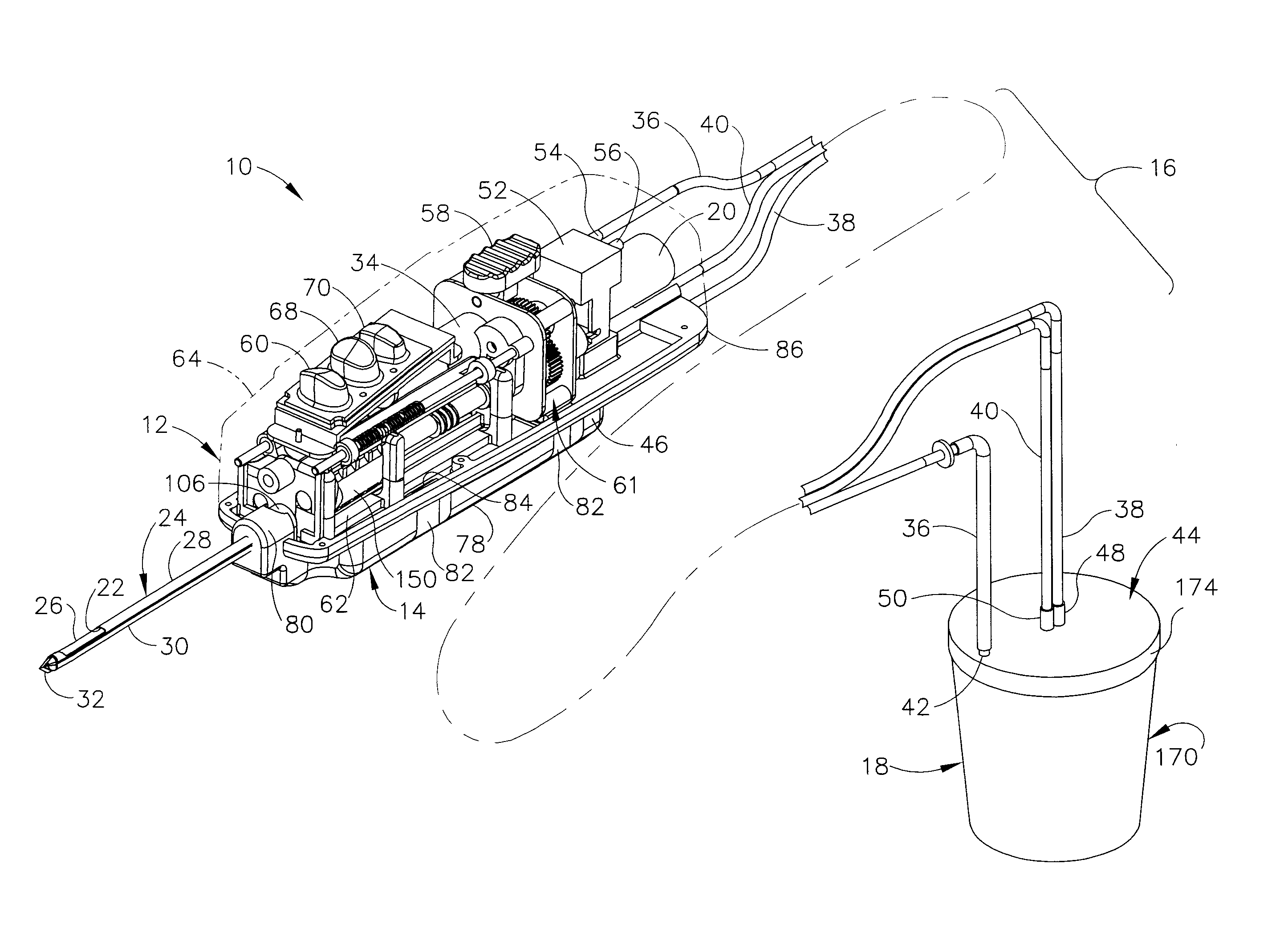
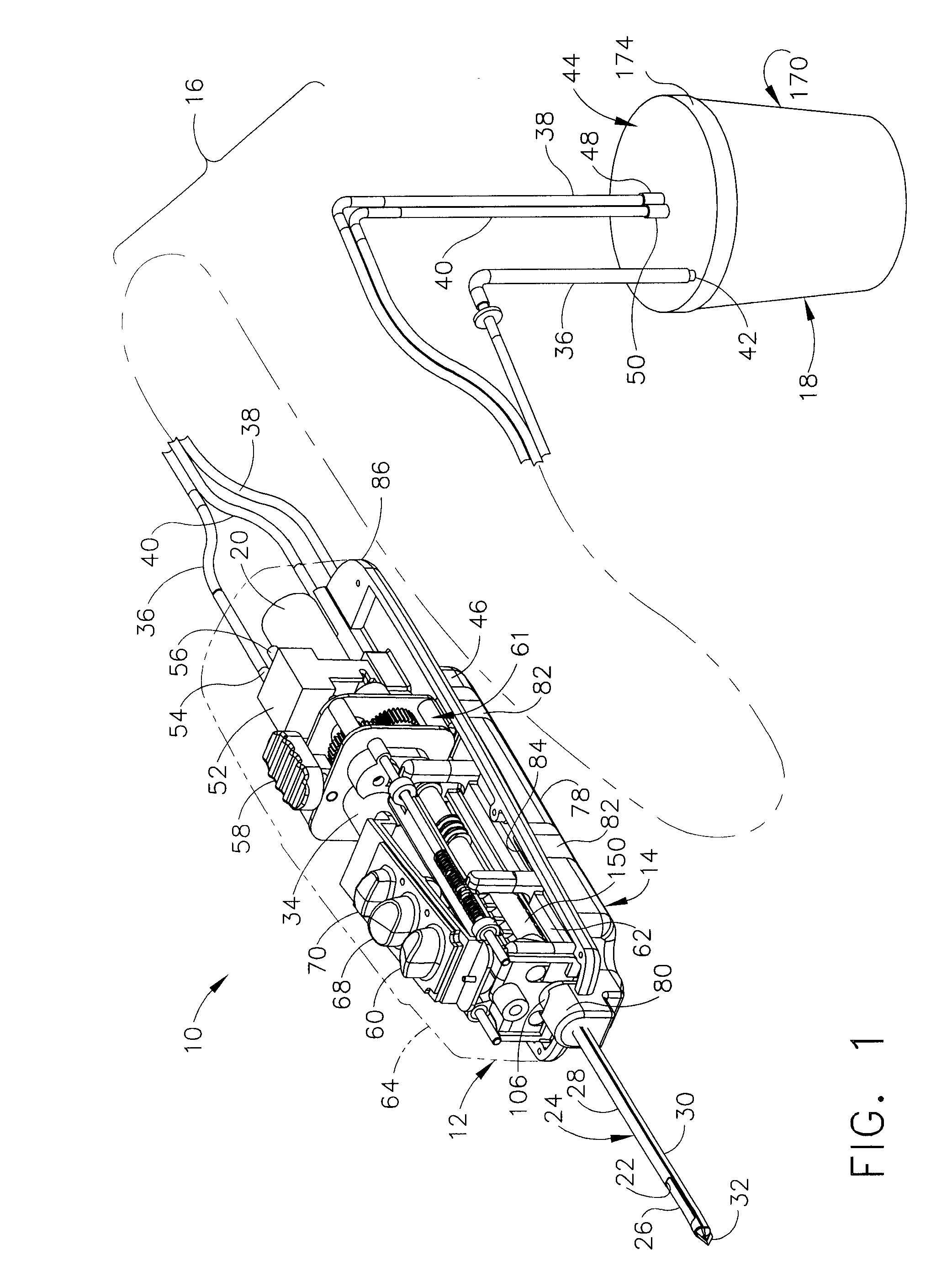
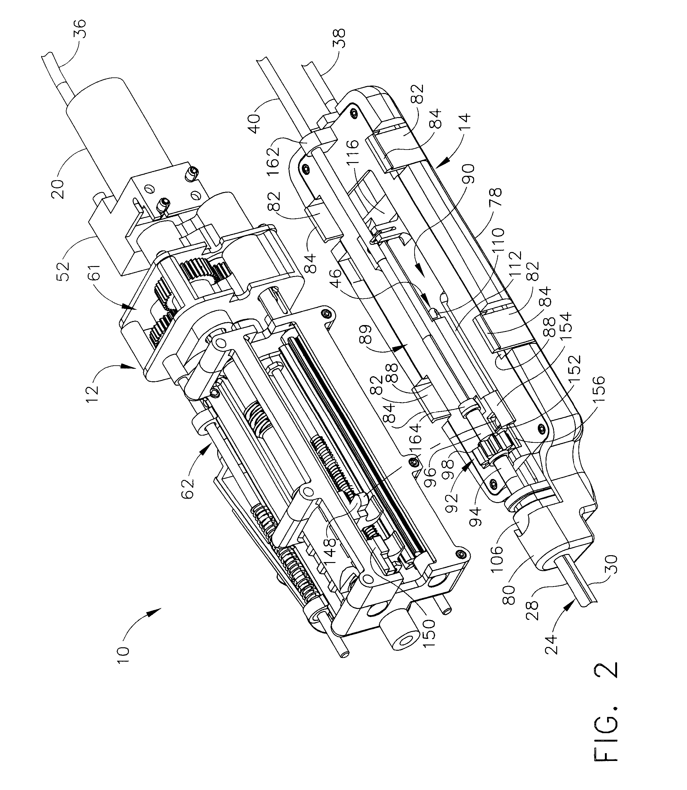
[0022] Turning to the Drawings, wherein like numerals denote like components throughout the several views, in FIG. 1, a biopsy device 10 includes a reusable handpiece 12 and a disposable probe assembly 14. A self-contained biopsy system 16 is formed by attaching a replaceable biopsy sample and fluid capturing canister 18. The canister 18 is generally sized to accommodate comfortably a volume of fluid that would be extracted, including saline flushing during a biopsy procedure with sufficient internal volume as well to hold biopsy tissue samples 19 (FIG. 6). As such, biohazards associated with bodily tissue and fluids are mitigated in that all such materials are readily transported from a biopsy suite for pathology assessment without the necessity of on-site access.

[0023] Tissue is drawn by vacuum assistance generated by vacuum pump 20 integral to the reusable handpiece 12 into a side aperture 22 of a probe cannula 24 of the disposable probe assembly 14. The pneumatic vacuum assista...


PUM


Abstract

A biopsy device is provided for obtaining a tissue sample, such as a breast tissue biopsy sample. The biopsy device includes a disposable probe assembly with an outer cannula having a distal piercing tip, a cutter lumen, and a cutter tube that rotates and translates past a side aperture in the outer cannula to sever a tissue sample. The biopsy device also includes a reusable handpiece with an integral motor and power source to make a convenient, untethered control for use with ultrasonic imaging. The reusable handpiece incorporates a probe oscillation mode to assist when inserting the distal piercing tip into tissue. An integral vacuum motor assists prolapsing tissue for effective severing as well as facilitating withdrawal of the tissue samples and bodily fluids from the biopsy site into a detachable, self-contained canister for transporting the separated biopsy samples and fluid for pathology assessment, avoiding biohazards in a clinical setting.

Description

CROSS REFERENCE TO RELATED APPLICATIONS [0001] The present application is a continuation-in-part of the co-pending and commonly-owned U.S. patent application Ser. No. 10 / 953,834, “BIOPSY APPARATUS AND METHOD” to Hibner et al., filed 29 Sep. 2004 (U.S. Pat. Publ. No. 2006 / 0074345) (END-5469USNP), the disclosure of which is hereby incorporated by reference in its entirety.FIELD OF THE INVENTION [0002] The present invention relates in general to biopsy devices, and more particularly to biopsy devices having a cutter for severing tissue, and even more particularly to biopsy devices for multiple sampling with a probe remaining inserted. BACKGROUND OF THE INVENTION [0003] When a suspicious tissue mass is discovered in a patient's breast through examination, ultrasound, MRI, X-ray imaging or the like, it is often necessary to perform a biopsy procedure to remove one or more samples of that tissue in order to determine whether the mass contains cancerous cells. A biopsy may be performed usi...

Claims


Application Information

Patent Timeline
Patent Type & Authority Applications(United States)
IPC IPC(8): A61B10/02
CPCA61B10/0275A61B2010/0225A61B2010/0208A61B10/0283
Inventor HIBNER, JOHN
Owner DEVICOR MEDICAL PROD
Why Eureka
Try Eureka