
Systems and methods for image presentation for medical examination and interventional procedures

Inactive Publication Date: 2010-05-13
FUJIFILM SONOSITE
View PDF10 Cites 66 Cited by
  • Summary
  • Abstract
  • Description
  • Claims
  • Application Information

AI Technical Summary

Benefits of technology

[0013]Embodiments of the invention additionally or alternatively restrict the numbers and types of images which may be displayed. According to embodiments of the invention, the degrees of freedom for image plane rotation about various axes of a multi-dimensional volume, such as when selecting an image plane for generating images. For example, although 6 degrees of freedom (e.g., bidirectional rotation about the X axis, bidirectional rotation about the Y axis, and bi-directional rotation about the Z axis) are often available with respect to a 3D volume, embodiments of the present invention impose restrictions to the degrees of freedom with respect to image plane selection. According to an embodiment of the invention, the ability to generate MPR images from a multi-dimensional image volume are limited to cross-sectional images generated along a particular axis, arc, etc. The use of image plane freedom limitations according to embodiments of the invention synergistically is accompanied by a simplified user interface. For example, where the ability to generate MPR images is limited to cross-sectional images generated along an axis or arc, embodiments of the invention may implement a relatively simple bidirectional control, such as left and right buttons, spinner knob, single axis joystick, etc. Moreover, the use of image plane freedom limitations according to embodiments of the invention preferably facilitates a user's ability to obtain an image or images needed or desired for a particular task or use, as well as preventing a user from accidentally failing to obtain a needed or desired image.

Problems solved by technology

However, the present inventors have discovered that when imaging less recognizable structure and / or utilizing images for interventional procedures, such image display freedom can lead to an inability to interpret the image and confusion by the person examining the image.
Where a technician, such as a physician, is attempting an interventional procedure, such as inserting a needle or catheter in a patient to achieve a precise placement, such freedom in displaying the image can result in confusion and an inability to determine the correct movements to be made.
Accordingly, the use of 3D and 4D medical imaging has heretofore been limited in its applicability.

Method used


Image

  • Systems and methods for image presentation for medical examination and interventional procedures
  • Systems and methods for image presentation for medical examination and interventional procedures
  • Systems and methods for image presentation for medical examination and interventional procedures

Examples


Embodiment Construction

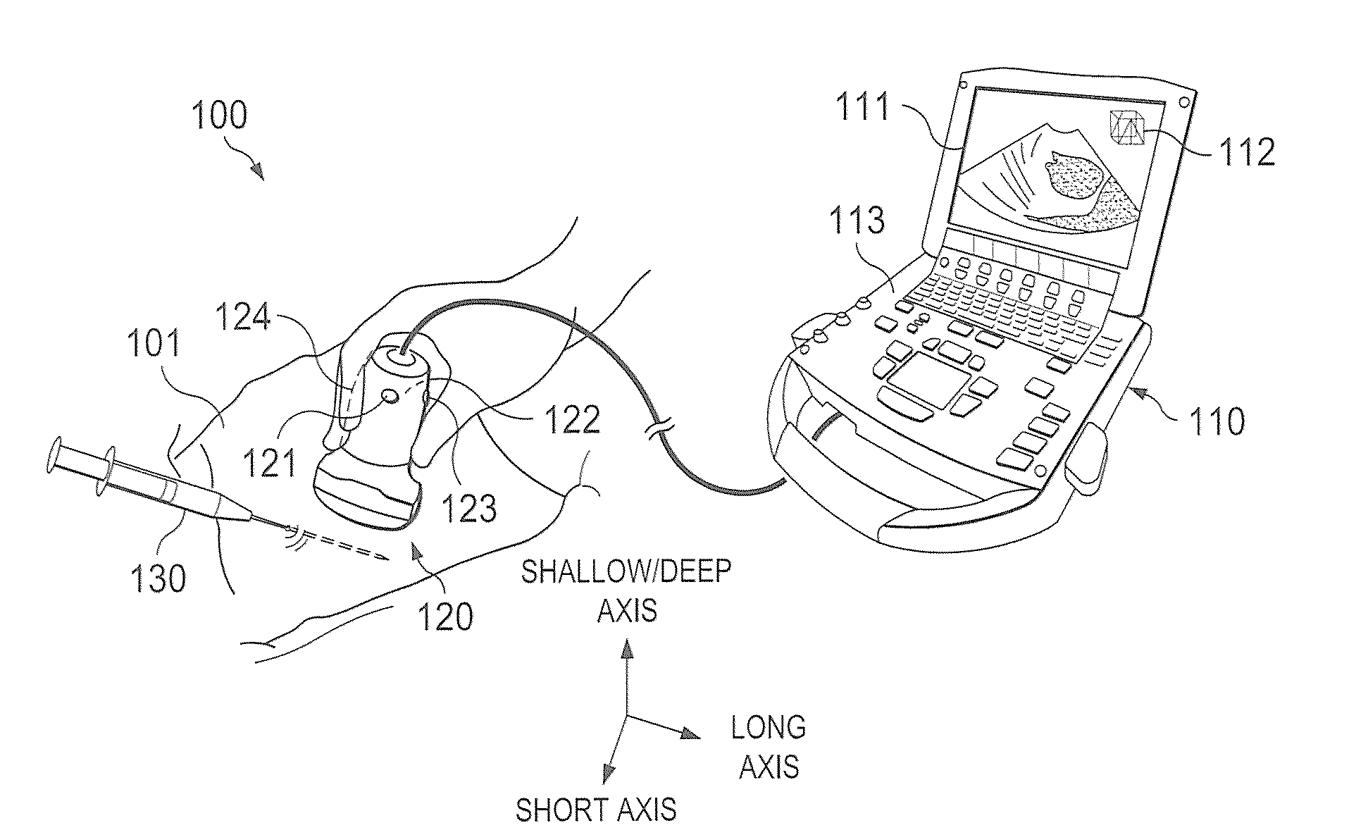
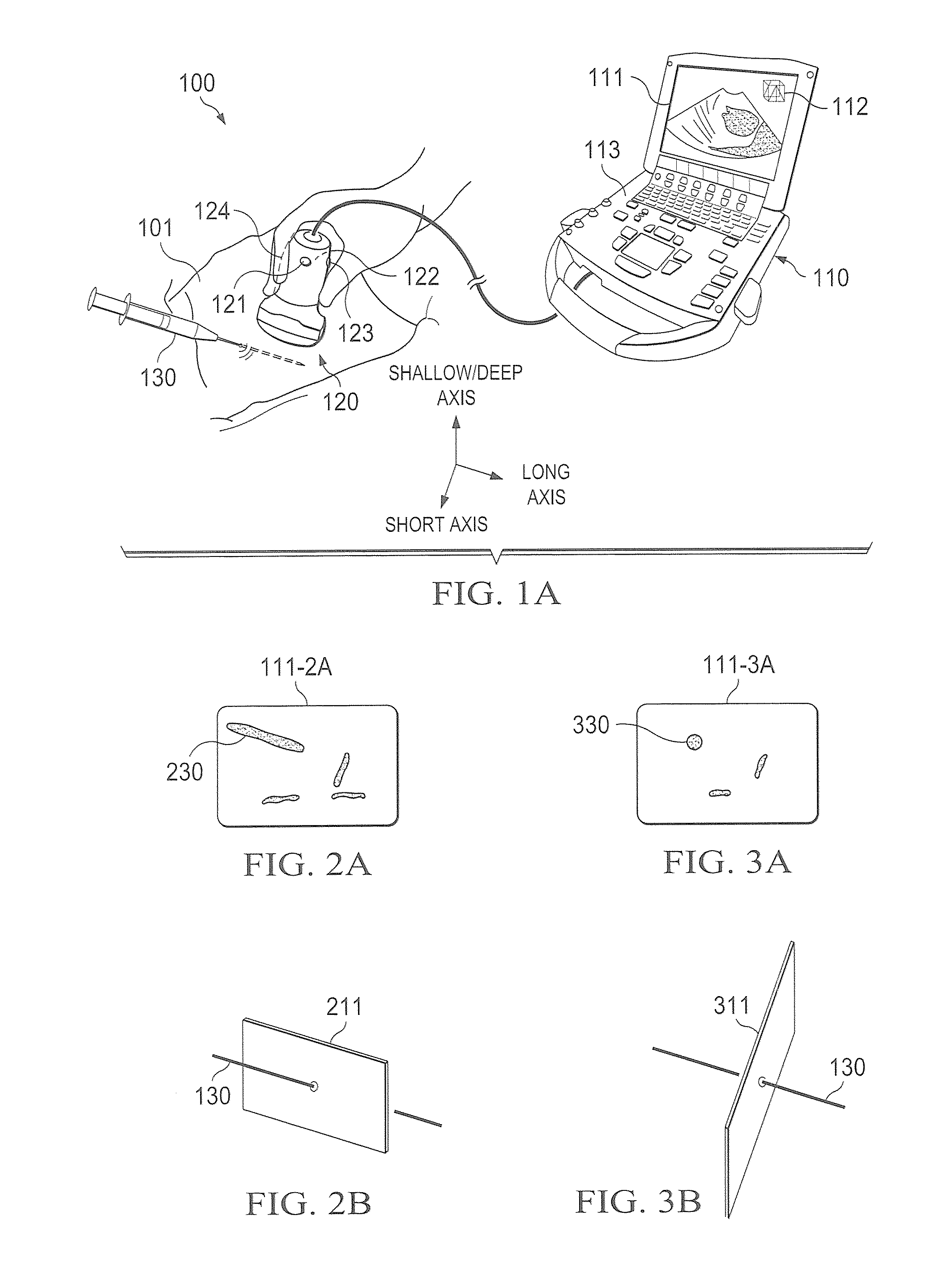
[0023]Directing attention to FIG. 1A, a system adapted according to embodiments of the invention is shown as system 100. System 100 may, for example, comprise a diagnostic ultrasound system operable to provide 2D and / or 3D images from a multi-dimensional (e.g., 3D and / or 4D) volume dataset. Although embodiments of the invention are described herein with reference to ultrasound imaging technology, in order to aid the reader in understanding the invention, it should be appreciated that the concepts of the present invention are not limited in applicability to ultrasound imaging. For example, embodiments of the present invention may be implemented with respect to fluoroscope systems, X-ray imaging systems, ultrasound imaging systems, CT imaging systems, MRI systems, positron emission tomography (PET) imaging systems, and the like.

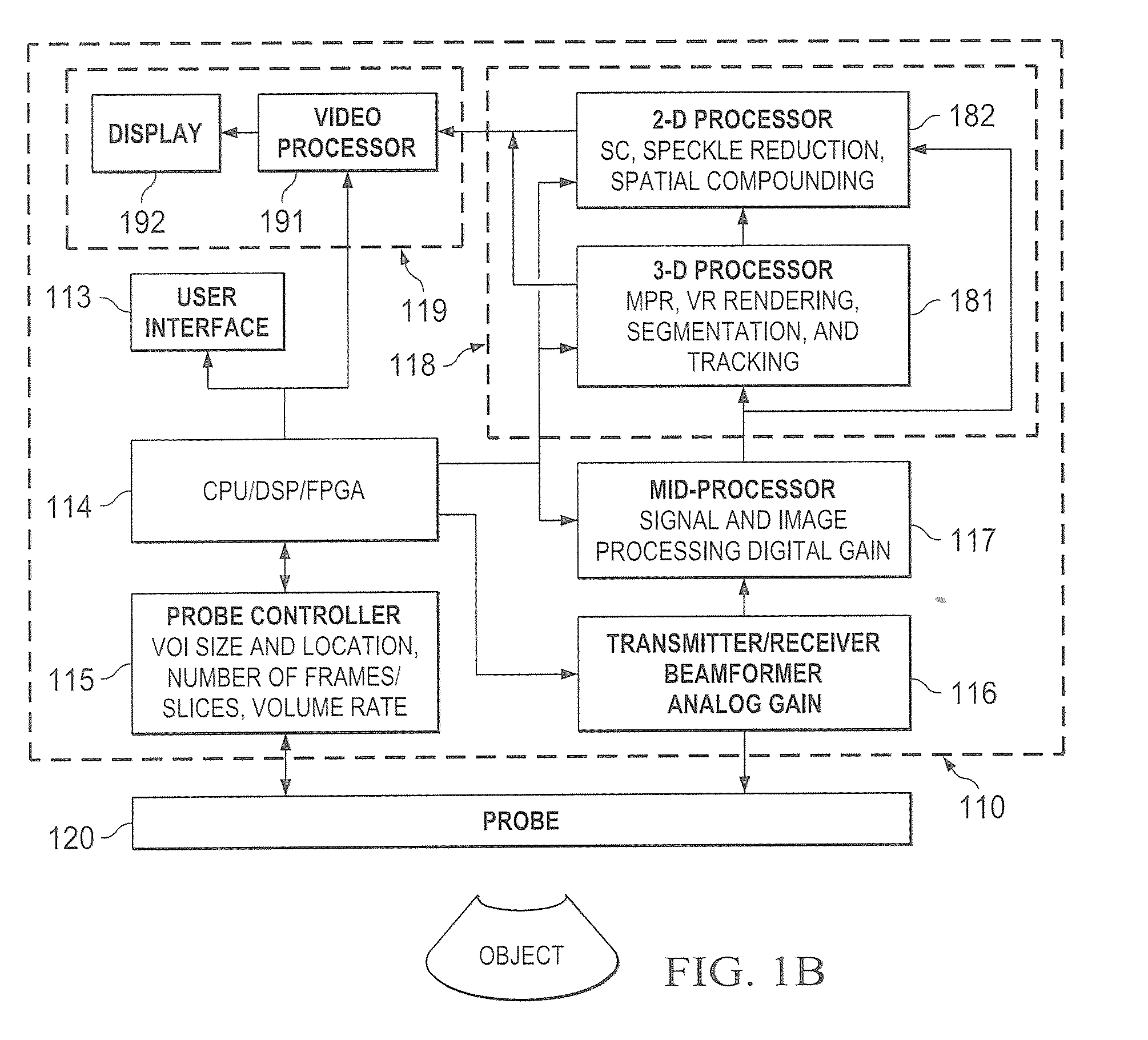
[0024]System 100 of the illustrated embodiment includes system unit 110 and transducer 120 coupled thereto. System unit 110 preferably comprises a processor-ba...


PUM


Abstract

Systems and methods which provide image presentation for medical examination, interventional procedures, diagnosis treatment, etc. from multi-dimensional volume datasets are shown. Reference indicators, providing information with respect to the relationship of an image to the physical world, are preferably provided to aid a viewer in interpreting the image. Such reference indicators may be provided in the form of tool markers and corresponding image marker. Degrees of freedom provided with respect to image manipulation are preferably selectively constrained to facilitate interaction with an image or images. Embodiments of the invention may implement a relatively simple bidirectional control to facilitate a survey of an entire image volume. Image display conventions may be provided which present images in a particular orientation to facilitate user interpretation of the image.

Description

REFERENCE TO RELATED APPLICATIONS[0001]The present application is related to co-pending and commonly assigned U.S. patent application Ser. No. [Attorney Docket No. 65744-P044US-10805628] entitled “Systems and Methods to Identify Interventional Instruments,” filed concurrently herewith the disclosure of which is hereby incorporated herein by reference.TECHNICAL FIELD[0002]The invention relates generally to image presentation and, more particularly, to image presentation for medical examination, interventional procedures, diagnosis, treatment, etc.BACKGROUND OF THE INVENTION[0003]Various forms of imaging apparatus have been used extensively for medical applications. For example, fluoroscope systems, X-ray imaging systems, ultrasound imaging systems, computed tomography (CT) imaging systems, and magnetic resonance (MR) imaging (MRI) systems have been used for a number of years. For example, medical examination, interventional procedures, diagnosis, and / or treatment may be provided usin...

Claims


Application Information

Patent Timeline
IPC IPC(8): A61B8/00G06K9/00
CPCA61B8/00G06T19/00A61B8/483A61B8/461A61B8/463A61B8/466
Inventor MA, QINGLINPAGOULATOS, NIKOLAOS
Owner FUJIFILM SONOSITE
Learn More