Imaging method for microcalcification in tissue and imaging method for diagnosing breast cancer

a microcalcification and imaging method technology, applied in the field of imaging method for breast cancer and imaging method for diagnosing breast cancer, can solve the problems of less than 30% sensitivity of microcalcification image and huge challenge for all clinical practitioners, and achieve the effects of no speckle noise, high optical contrast and high ultrasonic resolution

Inactive Publication Date: 2011-06-16
NATIONAL TSING HUA UNIVERSITY
View PDF6 Cites 21 Cited by
  • Summary
  • Abstract
  • Description
  • Claims
  • Application Information

AI Technical Summary

Benefits of technology

[0008]The present invention is directed to an imaging method for microcalcification which displays microcalcification distribution by acquiring and overlapping a photoacoustic image of microcalcification and an ultrasonic image of tissue. The image acquired by the present invention, in comparison to images acquired by ultrasonic and X-ray mammography, has advantages in no speckle noises, higher optical contrast, higher ultrasonic resolution, and so on.

Problems solved by technology

However, the X-ray mammogram may display microcalcification but not other subtle structures, such as mammary ducts.
However, due to speckle noises present in the ultrasound image and relative low contrast between the breast tissue and microcalcification, the sensitivity of the microcalcification image is less than 30%.
Therefore, it has been a huge challenge for all clinical practitioners to find out the position of suspected tumor and microcalcification found in X-ray mammogram using an ultrasound system.

Method used


Image

  • Imaging method for microcalcification in tissue and imaging method for diagnosing breast cancer
  • Imaging method for microcalcification in tissue and imaging method for diagnosing breast cancer
  • Imaging method for microcalcification in tissue and imaging method for diagnosing breast cancer

Examples


Embodiment Construction

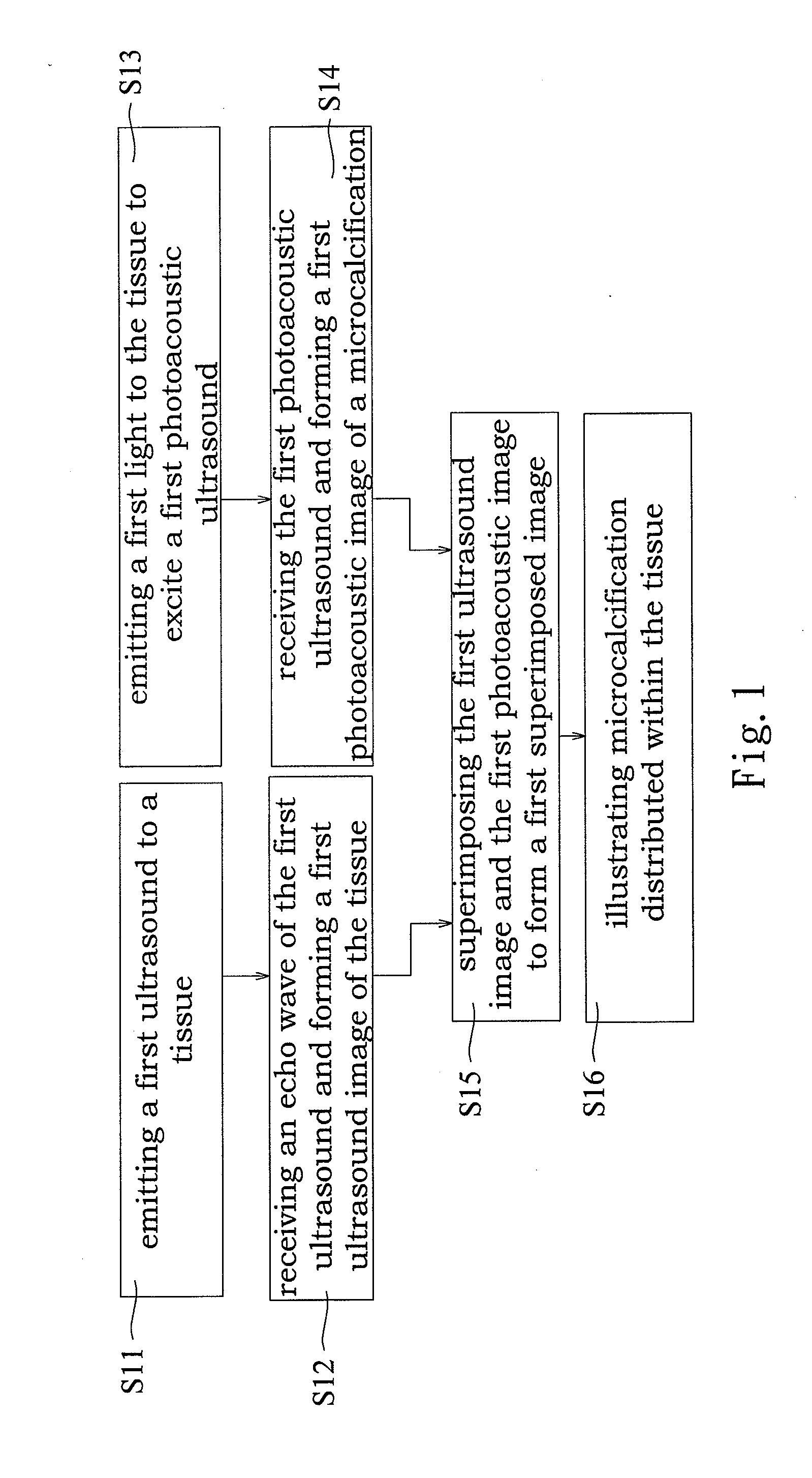
[0022]The present invention is directed to a superimposed image generated from a photoacoustic image of microcalcification on an ultrasound image of the tissue. The superimposed image is used for illustrating microcalcification distributed within the tissue.

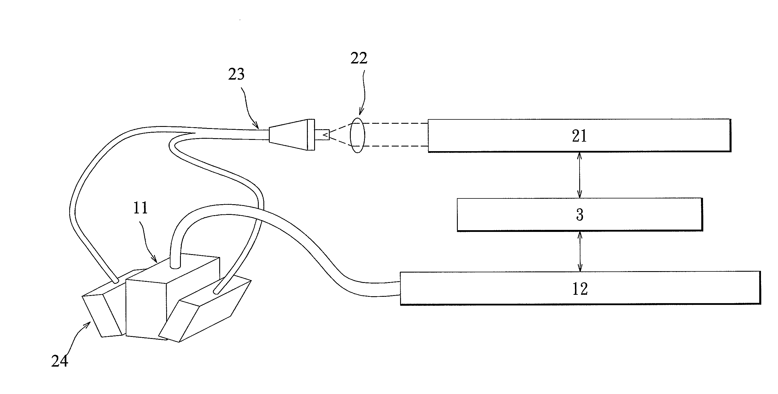
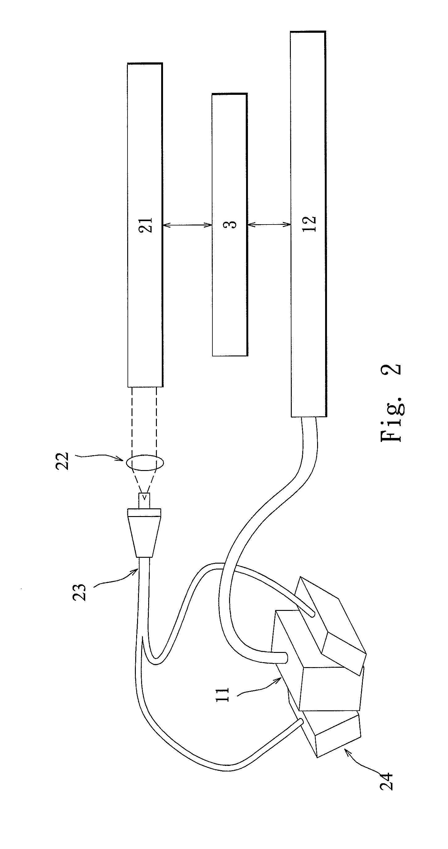
[0023]Refer to FIG. 1 and FIG. 2, where FIG. 1 is a flow chart illustrating an imaging method for microcalcification according to an embodiment of the present invention, and FIG. 2 is a schematic diagram illustrating an integrated system of photoacoustic imaging and ultrasound imaging according to an embodiment of the present invention.

[0024]First, Steps S11 and S12 are performed to obtain an ultrasound image of ROI (Region of Interest) tissue. Step S11 includes emitting a first ultrasound to a tissue. The ultrasound may be generated with an ultrasound array transducer 11. Short electrical pulses generated with the ultrasound array transducer 11 result in the ultrasound at the desired frequency. The ultrasound array transducer 11...


PUM


Abstract

An imaging method for microcalcification displays microcalcification distribution by acquiring and overlapping a photoacoustic image of microcalcification and an ultrasonic image of tissue. The image acquired by the present invention, in comparison to images acquired by ultrasonic and X-ray mammography, has advantages in no speckle noises, higher optical contrast, higher ultrasonic resolution, and so on. The present invention also has advantage in safety by adopting a light source having no ionizing radiation. An imaging method for diagnosing breast cancer is also herein disclosed.

Description

BACKGROUND OF THE INVENTION[0001]1. Field of the Invention[0002]The present invention relates to an imaging method for microcalcification in tissue and imaging method for diagnosing breast cancer, particularly to an imaging method for microcalcification in tissue and imaging method for diagnosing breast cancer by acquiring and superimposing a photoacoustic image of microcalcification and an ultrasonic image of tissue.[0003]2. Description of the Prior Art[0004]Microcalcification in breast has been one of the important indicators for early diagnosis for breast cancer and the location and distribution of microcalcification is also an important indicator for distinguishing malignant breast tumor. Therefore, detection of microcalcification in the breast is important for early diagnosis of breast tumor. X-ray mammogram is the most economical and effective screening tool among all and the only approved screening tool for breast cancer in many countries, including the United States, around ...

Claims


Application Information

Patent Timeline
Patent Type & Authority Applications(United States)
IPC IPC(8): A61B8/14
CPCA61B5/0095A61B8/06A61B5/0091A61B8/13A61B8/5238A61B8/0825A61B5/7425
Inventor LI, MENG-LINHSIAO, TSAI-CHUCHEN, SHIN-CHEHCHENG, YAO-YUWANG, PO-HSUNFAN, CHIH-TAI
Owner NATIONAL TSING HUA UNIVERSITY
Why Eureka
Try Eureka