Diagnostic imaging device for medical use

a diagnostic imaging and medical technology, applied in the direction of instruments, patient positioning for diagnostics, applications, etc., can solve the problems of not obtaining ct images from the x-ray ct device for diagnostic purposes, exposing the examination subject to x-rays, etc., to avoid unnecessary exposure to x-rays, improve diagnostic accuracy, and improve safety

Inactive Publication Date: 2006-12-26
SHIMADZU CORP
View PDF5 Cites 47 Cited by
  • Summary
  • Abstract
  • Description
  • Claims
  • Application Information

AI Technical Summary

Benefits of technology

[0011]The control device controls the PET data collection unit and PET image reconstruction unit to collect PET data and reconstruct PET images for an examination region of the examination subject and displays the obtained PET images, and then prompts the doctor to enter an input indicating whether to advance to CT data collection and CT image reconstruction processes or not. The doctor can enter either an instruction to advance to CT data collection and CT image reconstruction if the doctor judges that there is an indication of possible cancer, or an instruction not to advance to CT scanning if the doctor judges that there is no indication of cancer upon observation of the displayed PET images. Because the PET scanning and PET image displays are done first and then the doctor who has seen the images is prompted to enter a decision whether to advance to CT scanning, the doctor is able to concentrate on the diagnosis of cancer by means of the PET images and is helped on the decision process of whether to advance to the next step of CT examination. The whole process is made easier for the doctor, who can order CT scanning only when there is a suspicion for cancer. This avoids the risk of unnecessary exposure to X-rays while taking advantage of the PET examination which provides an efficient means to detect cancer early.
[0012]If the control device can further make a determination as to whether the pixel values of the PET images obtained from the preliminary PET scanning exceeds a specified threshold value, this may help the doctor in making a close observation of the PET images to see if there are any regions where cancers can be suspected, supporting the doctor's diagnosis and making the job of early detection of cancers easier, safer and more secure.

Problems solved by technology

However, the examination using an X-ray CT device causes a problem of exposing the examination subject to X-rays.
Moreover, although a system has been disclosed in Laid open patent application H7-20245 (patent document 1) in which a common examination table is used for a PET device and an X-ray CT device, such a system uses data obtained from the X-ray CT device for the absorption compensation of the PET data, and does not obtain CT images from the X-ray CT device for diagnostic purposes.

Method used


Image

  • Diagnostic imaging device for medical use
  • Diagnostic imaging device for medical use
  • Diagnostic imaging device for medical use

Examples


embodiment 1

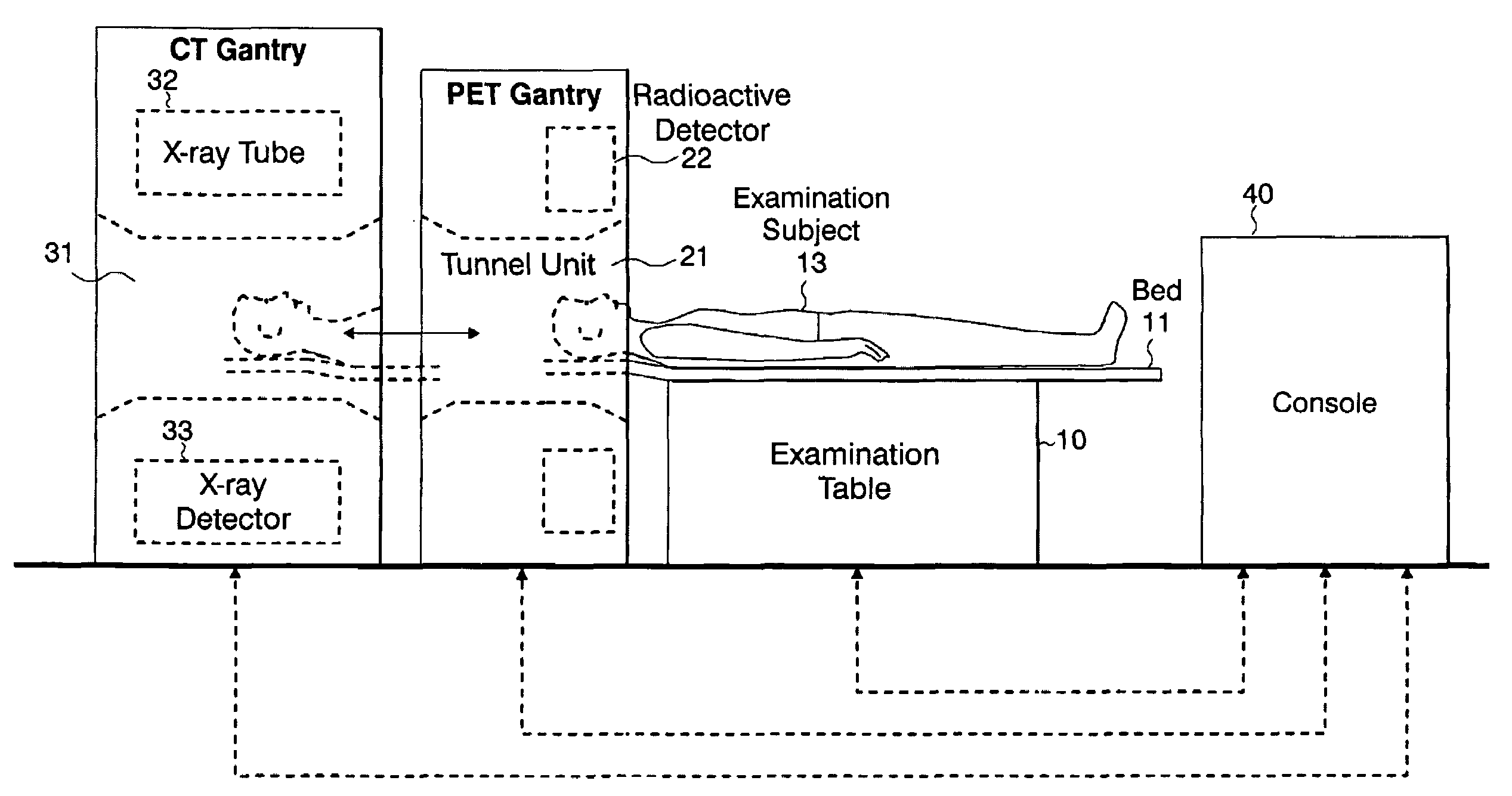
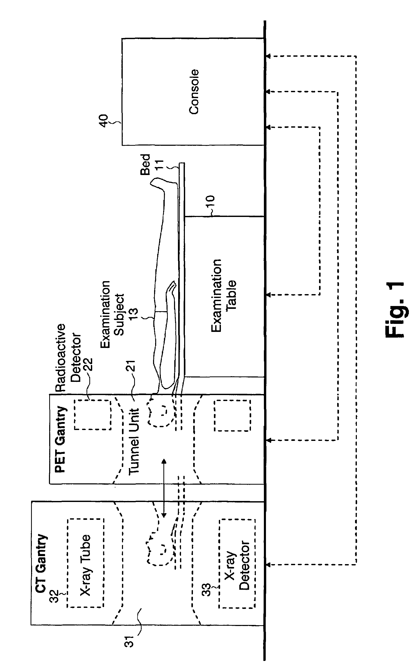
[0016]As shown in FIG. 1, the diagnostic imaging device for medical use according to this invention comprises an examination table 10, a PET gantry 20, a CT gantry 30, and a console 40. The PET gantry 20 constitutes the PET data collection unit, having a tunnel unit 21 in the center and radiation detectors 22 arranged like a ring around the tunnel unit 21. An examination subject 13 administered with a radio active drug is inserted into the tunnel unit 21, and the radiation detectors 22 arranged like a ring around it detects radiations released from the radiating drug existing in the examination subject 13. Two gamma rays released from a positron emitting nuclide at a time in 180° directions opposite to each other are detected as they enter simultaneously into two diametrically apart radiation detectors 22, which are arranged like a ring, and the detected data are collected in such a way that the presence of the radioactive drug is detected on the line connecting the two radiation de...


PUM


Abstract

A diagnostic imaging device is provided to detect cancers in early stages with a high degree of accuracy while minimizing the risk of X-ray exposure that can be caused by the X-ray CT examination, taking advantage of the PET examination which provides a means of efficient early detection of cancers while minimizing the risk of radiation exposure. A PET gantry 20 and a CT gantry 30 are arranged sequentially relative to a common examination table 10, a control unit provided in a console 40 controlling the examination table 10, the PET gantry 20, and the CT gantry 30, and an examination subject 13 laid on a top board 11 of the examination table 10 is inserted into a tunnel 21 of the PET gantry 20 for collecting PET data in order to reconstruct a PET image, display the PET image on an image monitor provided in the console 40, allow a doctor to make a determination as to whether the examination subject 13 should be further subjected to a CT examination by sending it into a tunnel 31 of the CT gantry 30, and allow necessary actions to be taken in accordance with the determination.

Description

INCORPORATION BY REFERENCE[0001]The present application claims priority under 35 U.S.C. §119 to Japanese Patent Application No. 2003-358014 filed on Oct. 17, 2003. The content of the application is incorporated herein by reference in its entirety.FIELD OF THE INVENTION[0002]The invention relates to a diagnostic imaging device for medical use, in particular, a diagnostic imaging device for medical use suitable for early detection of cancers.DESCRIPTION OF THE RELATED ART[0003]It is extremely important to detect cancers in early stages for their treatments. Among various kinds of methods and devices known for early detections of cancers, Positron Emission Tomography (PET) examination is considered one of the most promising methods because it can detect cancerous cells while they are quite small and does not cause any radiation exposure problem.[0004]The PET examination detects a distribution image (cross-sectional image) of positron emitting nuclides on a particular cross section of a...

Claims


Application Information

Patent Timeline
Patent Type & Authority Patents(United States)
IPC IPC(8): G01T1/164A61B5/05A61B6/00G01T1/161A61B6/03G01T1/29
CPCA61B6/032A61B6/037A61B6/5235G01T1/1611G01T1/2985A61B6/03
Inventor AMANO, MASAHARU
Owner SHIMADZU CORP
Why Eureka
Try Eureka