Attenuation correction for nuclear medical imaging scanners with simultaneous transmission and emission acquisition

a nuclear medical imaging and emission acquisition technology, applied in the field of attenuation correction of emission data, can solve problems such as emission contamination (ec) of transmission data

Inactive Publication Date: 2008-12-16
SIEMENS MEDICAL SOLUTIONS USA INC
View PDF5 Cites 11 Cited by
  • Summary
  • Abstract
  • Description
  • Claims
  • Application Information

AI Technical Summary

Problems solved by technology

Typically the recording of the transmission scan simultaneous with the recording of the emission scan will cause emission contamination (EC) of the transmission data.

Method used


Image

  • Attenuation correction for nuclear medical imaging scanners with simultaneous transmission and emission acquisition
  • Attenuation correction for nuclear medical imaging scanners with simultaneous transmission and emission acquisition
  • Attenuation correction for nuclear medical imaging scanners with simultaneous transmission and emission acquisition

Examples


Embodiment Construction

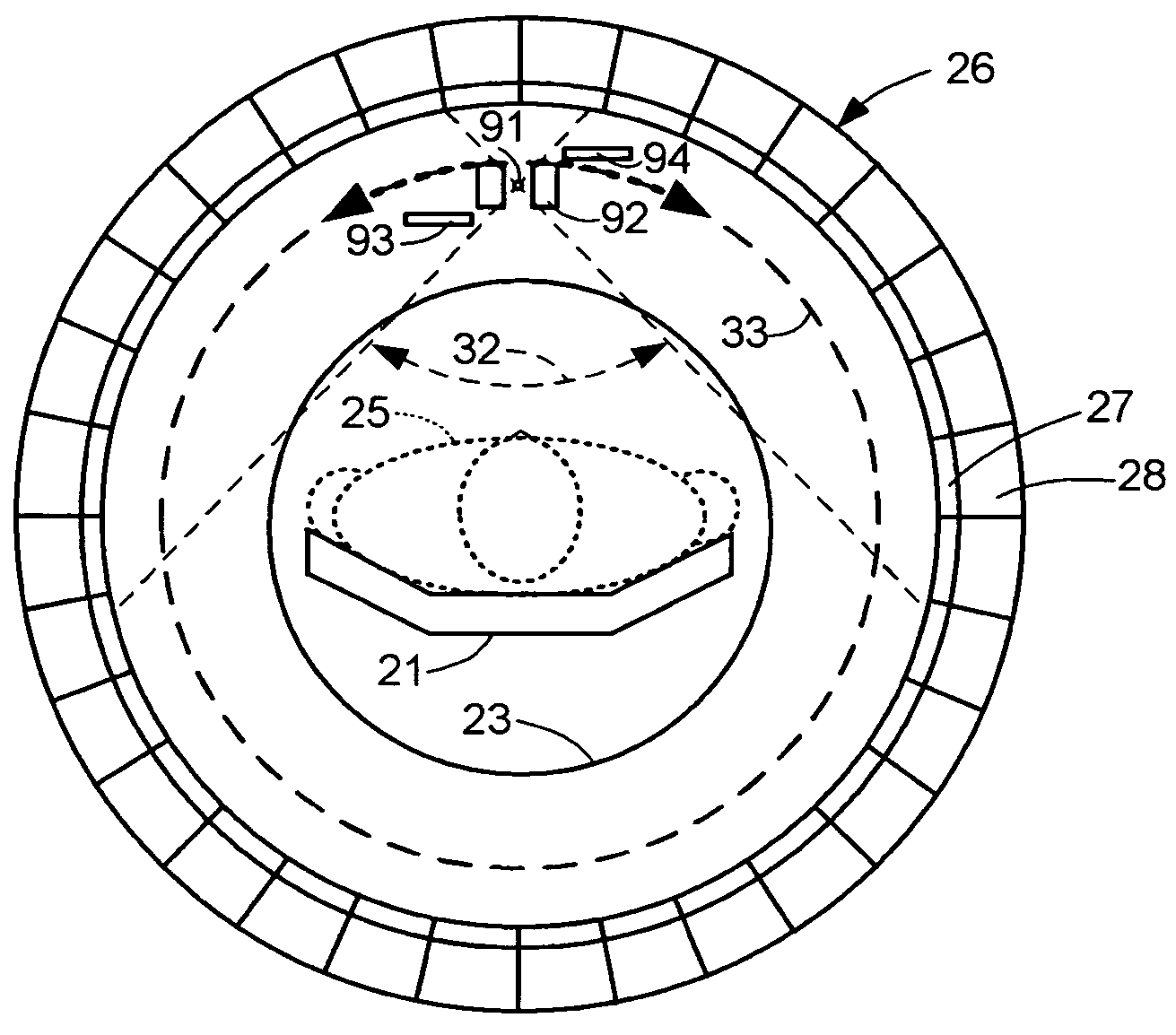
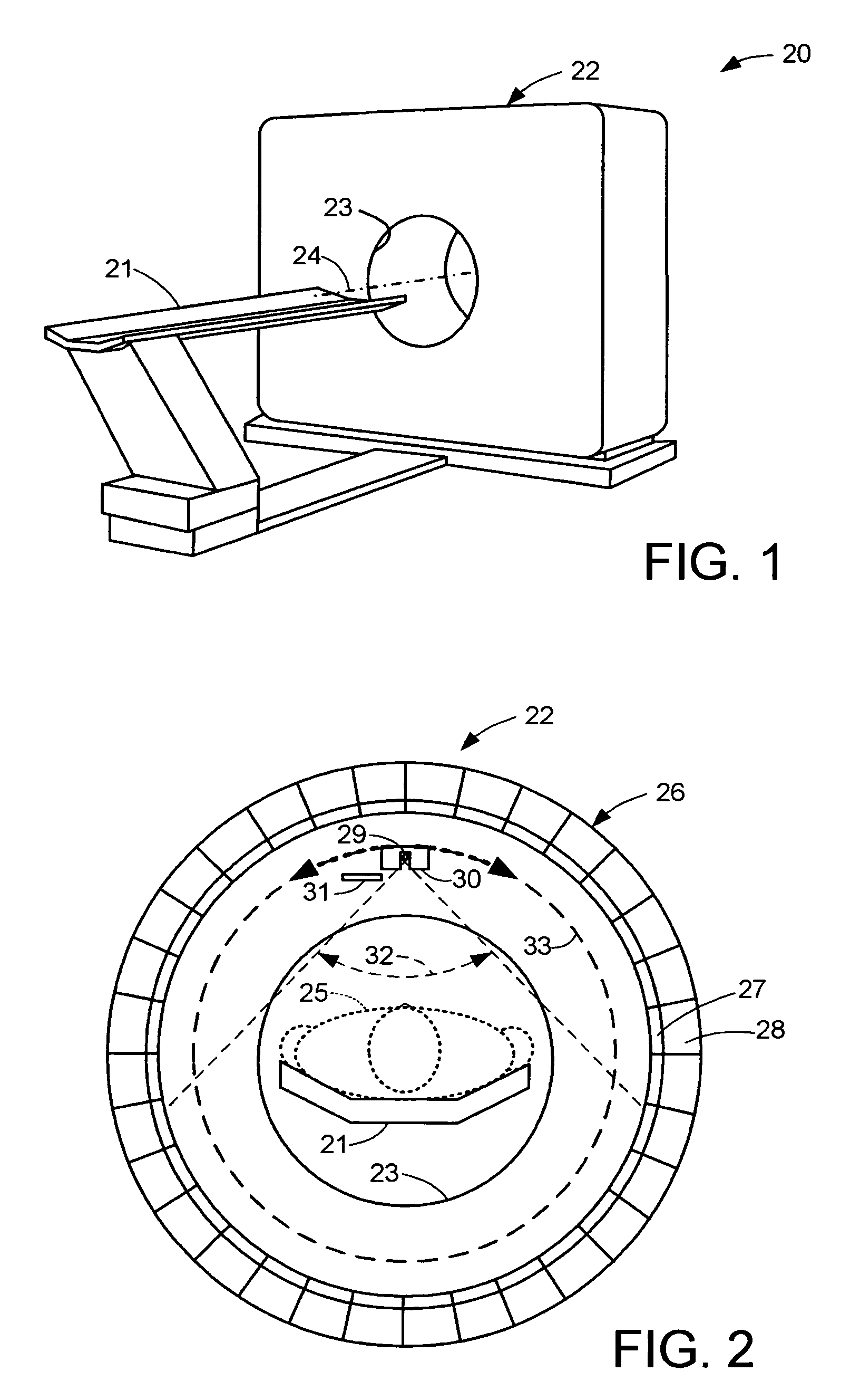
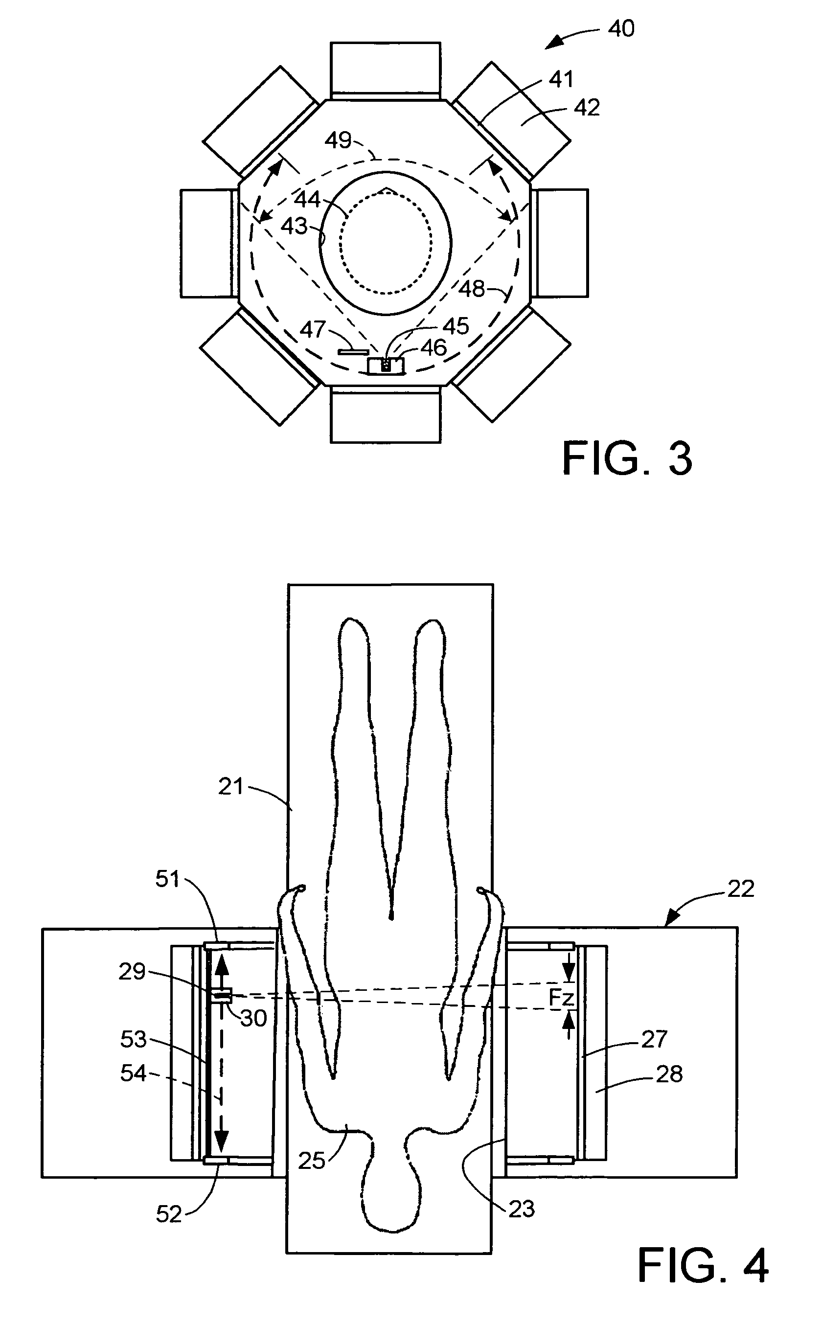
[0032]With reference to FIG. 1, there is shown a positron emission tomographic (PET) scanner generally designated 20. The scanner 20 includes a bed 21 for supporting a human patient (25 in FIG. 2) and a scanner assembly 22 having a cylindrical hole or tunnel 23 for receiving the patent. The bed 21 is aligned along an axis 24 of the tunnel 23 and is translated relative to the scanner assembly along this axial direction for scanning of the whole body of the patient.

[0033]As further shown in FIG. 2, when the patient 25 is received in the tunnel 23, the patient is surrounded by a gamma detector array 26 including panels of scintillating crystals 27 and photo-detectors 28. The scintillating crystals, for example, are made of Lutetium oxyorthosilicate (LSO) and are arranged in a cylindrical array having a spacing of about 4 mm between the centers of adjacent crystals in the axial and trans-axial directions. The photo-detectors, for example, are photomultiplier tubes or photodiodes. An 8×1...


PUM


Abstract

For patient transmission data acquired simultaneously with patient emission data, blank transmission data are acquired in the absence of the patient emission and therefore under count rate conditions different from the count rate conditions of the patient transmission data. To prevent the different count rate conditions from causing artifacts in reconstructed tomographic images, a correction is made for spatially varying count rate effects on the attenuation correction. For example, the blank scan data are adjusted according to the count rate at which the patient emission data are acquired, and the adjusted blank scan data and the patient transmission data are used for attenuation correction of the patient emission data used for reconstructing a tomographic image.

Description

BACKGROUND OF THE INVENTION[0001]1. Field of the Invention[0002]The present invention relates generally to nuclear medical (NM) imaging such as such as positron emission tomography (PET) and single photon emission computed tomography (SPECT), and more particularly to attenuation correction of emission data simultaneously acquired with transmission data.[0003]2. Description of the Background Art[0004]Medical imaging is one of the most useful diagnostic tools available in modern medicine. Medical imaging allows medical personnel to non-intrusively look into a living body in order to detect and assess many types of injuries, diseases, conditions, etc. Medical imaging allows doctors and technicians to more easily and correctly make a diagnosis, decide on a treatment, prescribe medication, perform surgery or other treatments, etc.[0005]There are medical imaging processes of many types and for many different purposes, situations, or uses. They commonly share the ability to create an image...

Claims


Application Information

Patent Timeline
Patent Type & Authority Patents(United States)
IPC IPC(8): G01T1/164
CPCG01T1/1617
Inventor PANIN, VLADIMIRCONTI, MAURIZIOCASEY, MICHAEL E.
Owner SIEMENS MEDICAL SOLUTIONS USA INC
Why Eureka
Try Eureka