Marker delivery system with obturator

a delivery system and biopsy technology, applied in the field of medical devices and methods, can solve the problems of blood clot risk, days or weeks before, may be removed, altered or obscured, etc., and achieve the effect of improving delivery and reducing the risk of clot formation

Active Publication Date: 2011-05-17
SENORX
View PDF8 Cites 38 Cited by
  • Summary
  • Abstract
  • Description
  • Claims
  • Application Information

AI Technical Summary

Benefits of technology

[0009]In one embodiment having features of the present invention the device includes an obturator which has an elongated shaft with a internal lumen, a proximal end, and a substantially sealed distal end which prevents or minimizes the backflow of body fluids, such as blood, though the lumen of the obturator. The substantially sealed distal end can be a penetratable membrane or may have petals or a duckbill-type valve which are configured to allow passage of one or more intracorporeal objects or a delivery tube with one or more intracorporeal objects therethrough while preventing or minimizing entry of body fluids into the inner lumen of the obturator. Preferably the obturator is configured to fit within a procedure cannula, e.g. a cannula of a biopsy device, for example, the cannula of SenoRx's EnCor™ Magnetic Resonance Imaging Breast Biopsy System. The cannula provides access to the desired location within the patient's body.
[0014]The devices, systems, and methods of the present invention offer improved delivery by minimizing the backflow of body fluids, such as blood, though the obturator lumen and thereby decreasing a risk of clot formation in the obturator. These and other advantages of the invention will become more apparent from the following detailed description when taken in conjunction with the accompanying drawings.

Problems solved by technology

This backflow of blood into the obturator creates a risk of blood clotting.
After the biopsy sample is taken, it may take several days or weeks before the results of the examination of the sample are obtained, and still longer before an appropriate treatment decision is reached.
However, radiographically imageable tissue features, originally detected in a mammogram, may be removed, altered or obscured by the biopsy procedure.

Method used


Image

  • Marker delivery system with obturator
  • Marker delivery system with obturator
  • Marker delivery system with obturator

Examples


Embodiment Construction

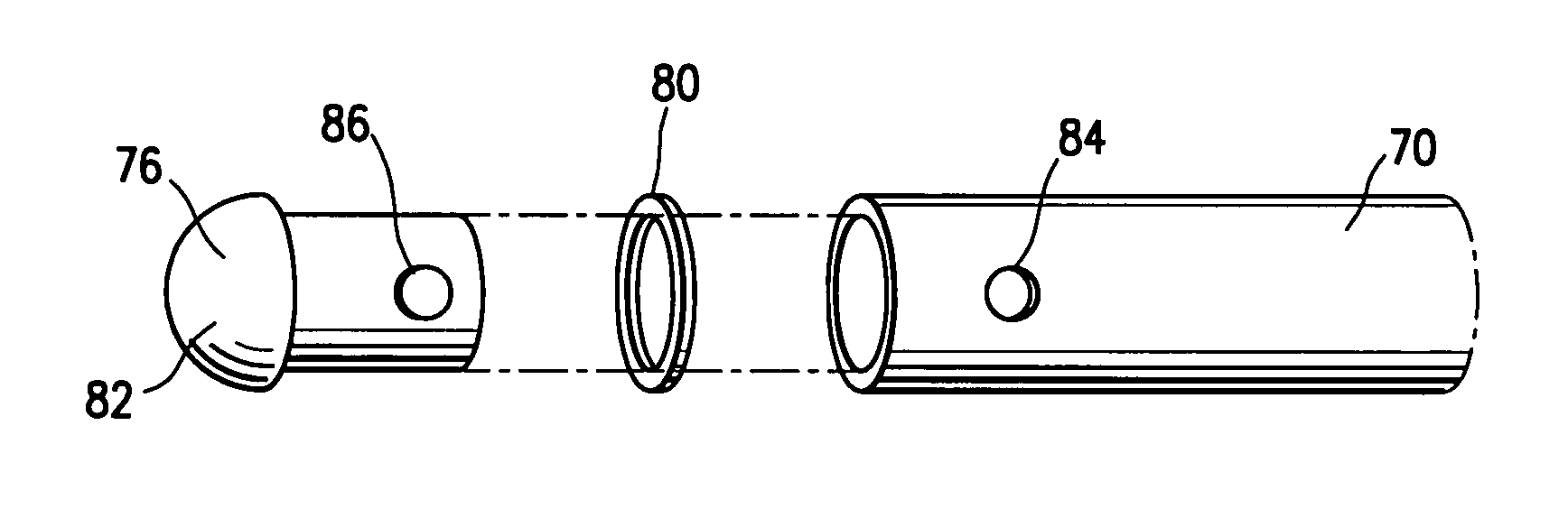
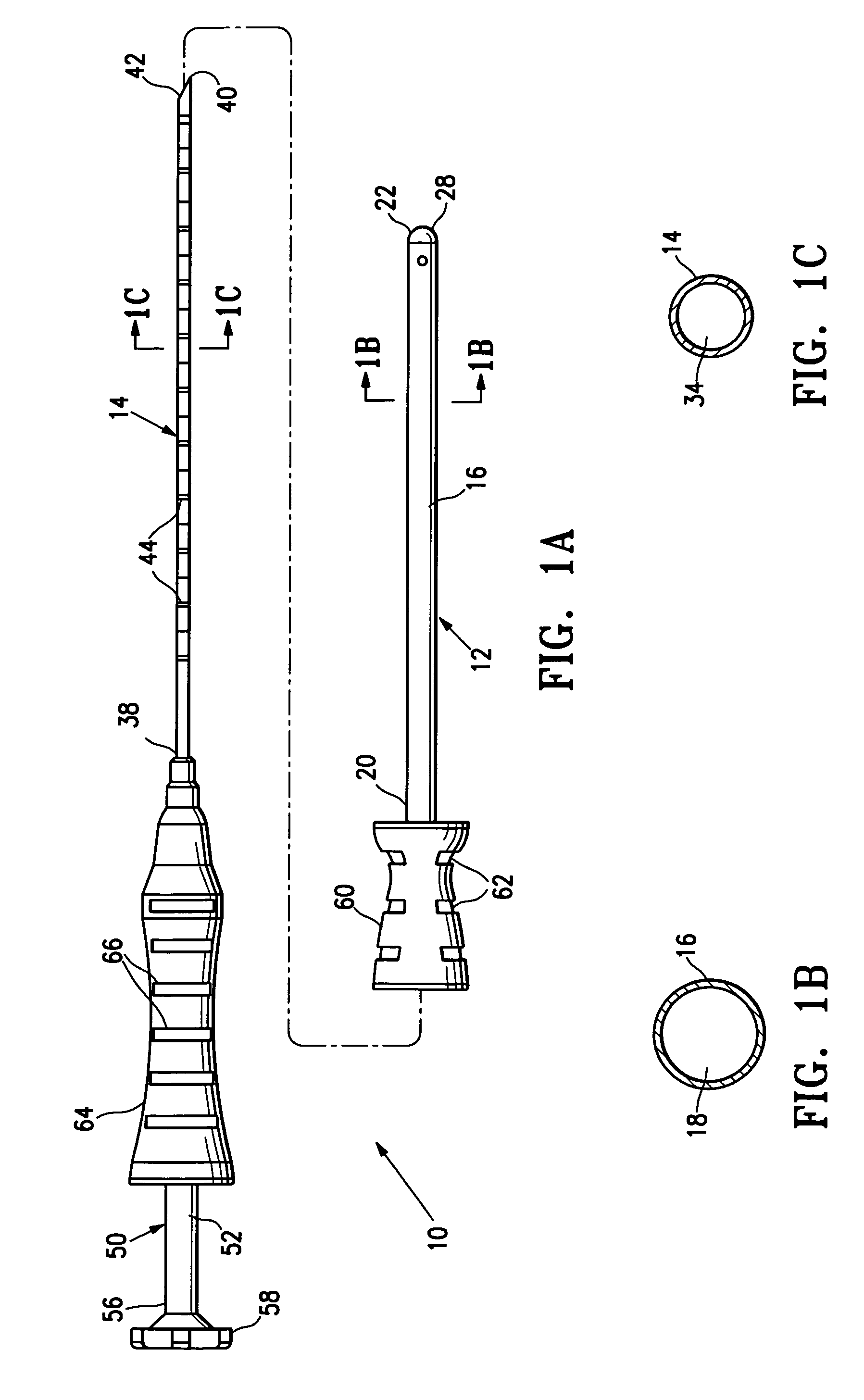
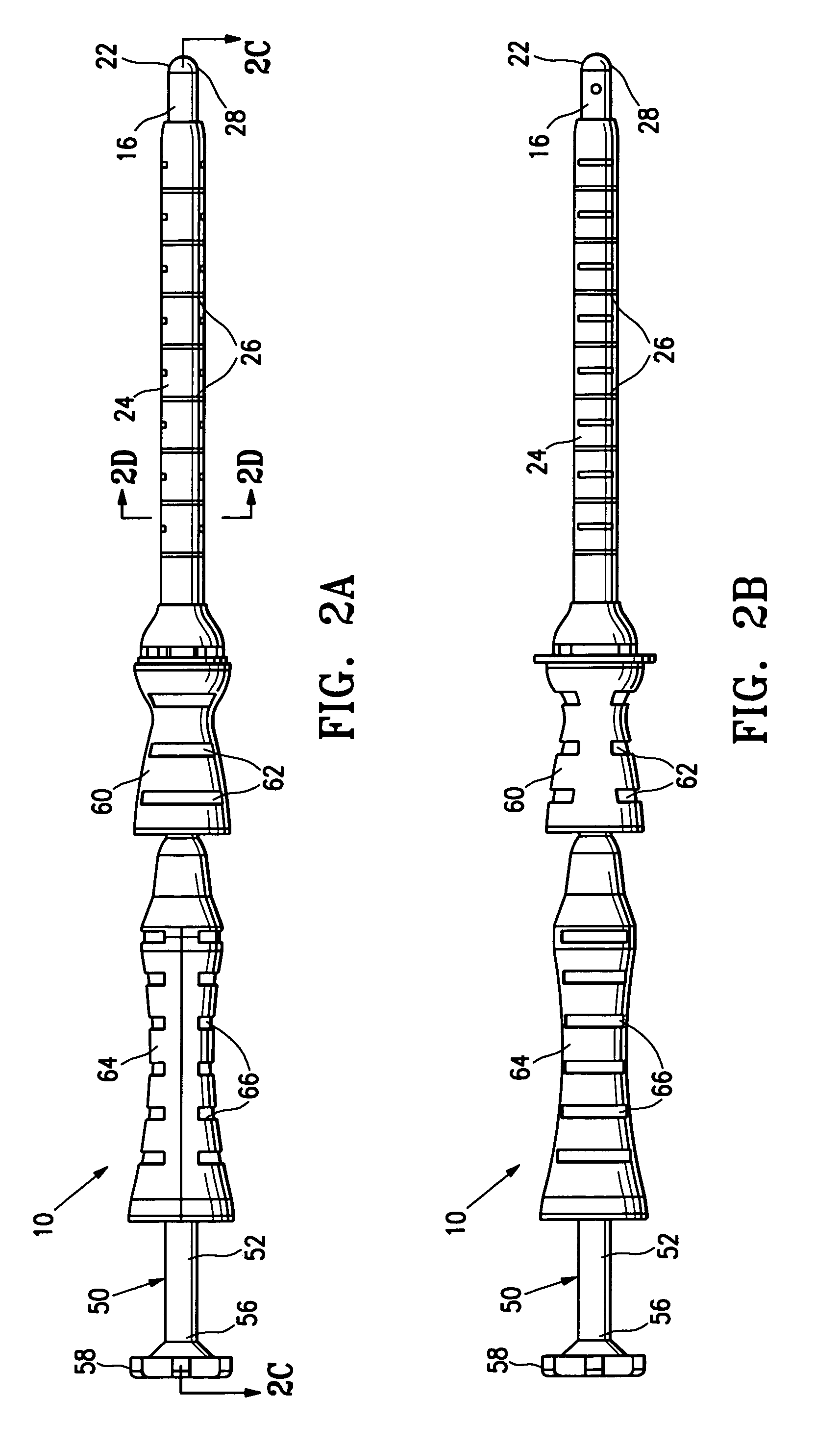
[0041]FIGS. 1A-2D shows an embodiment of a marker delivery device 10 having features of the invention including an obturator 12 and a marker delivery tubular shaft 14. The obturator 12 has an elongated shaft 16, an internal lumen 18, a proximal end 20 and a substantially sealed distal end 22. Preferably, as shown in FIGS. 2A-2D, the obturator 12 is configured to fit within a cannula 24 of a biopsy device, such as SenoRx's EnCor™ Magnetic Resonance Imaging (MRI) Breast Biopsy System. The cannula 24 provides access to the desired location within a patient's body. In some embodiments the cannula 24 includes depth markings 26 which indicate the distance which the obturator 12 has advanced within the patient's body.

[0042]The substantially sealed distal end 22 of the obturator 12 is configured to prevent or minimize the backflow of fluids, such as body fluids, through the internal lumen 18 of the obturator 12. Preferably the substantially sealed distal end 22 is formed of a penetratable m...


PUM


Abstract

A marker delivery device is described which has an obturator with an elongated shaft, an inner lumen, a proximal end, and a substantially sealed distal end. One or more tissue markers are deployed within the inner lumen of the elongated shaft of the obturator. Preferably, the tissue marker(s) is disposed within an inner lumen of a marker delivery tube which is disposed within the inner lumen of the elongated shaft of the obturator. The marker delivery tube has an opening for discharging the tissue markers into a body (e.g. biopsy) cavity. The distal tip of the marker delivery tube is configured to penetrate the substantially sealed distal end of the obturator so that tissue markers can be delivered while the obturator is in place within the body. Preferably, the obturator includes a detectable element capable of producing a relatively significant image signature during MRI.

Description

FIELD OF THE INVENTION[0001]This invention relates generally to the field of medical devices and methods. In particular, the invention relates to devices and methods for marking a biopsy site.BACKGROUND OF THE INVENTION[0002]In modern medical practice small tissue samples, known as biopsy specimens, are often removed from tumors, lesions, organs, muscles and other tissues of the body. Such removal of tissue samples may be accomplished by open surgical technique (i.e., removal of a small sample of tissue through a small surgical incision using a local anesthetic), or through the use of a specialized biopsy instrument such as a biopsy needle. After the tissue samples have been removed, they are typically subjected to diagnostic tests or examinations such as a) gross and microscopic examination to determine cytology and / or histology, b) biochemical analyses to determine the presence or absence of chemical substances which indicate certain disease states, c) microbiological culturing to...

Claims


Application Information

Patent Timeline
Patent Type & Authority Patents(United States)
IPC IPC(8): A61B5/05
CPCA61B17/3468A61B19/54A61B17/3498A61B19/38A61B2019/5487A61B2019/462A61B2019/5408A61B2019/5454A61B2017/00796A61B90/39A61B90/40A61B2090/062A61B2090/3908A61B2090/3954A61B2090/3987
Inventor LUBOCK, PAULQUICK, RICHARD
Owner SENORX
Why Eureka
Try Eureka