3D real-time tracking of human anatomy using combined kV and MV imaging

a real-time tracking and human anatomy technology, applied in the field of medical imaging devices and techniques, can solve the problems of increasing radiation exposure of additional kv imagers, not solving the problem, and tomographic rotation of kv or mv beams around the patient, so as to reduce radiation scatter

Active Publication Date: 2012-02-21
THE BOARD OF TRUSTEES OF THE LELAND STANFORD JUNIOR UNIV
View PDF9 Cites 24 Cited by
  • Summary
  • Abstract
  • Description
  • Claims
  • Application Information

AI Technical Summary

Benefits of technology

[0005]In contrast with hybrid systems, the present invention integrates kV and MV sources to overcome the disadvantages of prior systems. In one aspect, the present invention provides a method for the use of the MV radiation treatment beam information (captured using a detector) together with kV imaging for 3D tracking of internal features as a function of time. The method only requires the use of one kV source for full 3D localization, leading to dose reduction to the patient as compared with prior methods using multiple kV sources. The method also provides tracking in real time during treatment. In another aspect, the invention also provides a technique to maintain 3D tracking in the event that the fiducials / organs are blocked by the MV beam-defining aperture or high-density material such as bone. Additionally, a partial information method (PIM) allows continuous tracking of internal features in the event that the kV or MV beam is switched off for a specific amount of time. The PIM is beneficial when using intensity modulated radiation treatment (IMRT) and / or for reducing radiation scatter that adversely affects image quality.
[0006]Significantly, according to embodiments of the present invention the MV treatment beam is used in combination with kV imaging for the purpose of 3D geometric localization of internal features (e.g., organs and fiducials) during the radiation delivery process. The method provides several advantages over prior methods. These advantages may include, for example, lower total radiation exposure to the patient due to the treatment beam being used for geometric anatomy monitoring; near real-time monitoring of geometric target location to insure accurate radiotherapy procedures; and no need for significant hardware modification on treatment machines pre-equipped with kV and MV imaging devices. The method also provides the ability to use partial geometric and / or temporal information to compute the location of fiducials / organs in the event that the MV beam defining aperture blocks the view of the marker / organ on the MV imager or that the target moves behind dense objects as bone, thereby facilitating continuous tracking during IMRT delivery. It also provides the ability to use partial information to maintain full 3D tracking as a function of time even when either the kV beam or the MV beam is switched off for a limited amount of time, thereby allowing continuous tracking during IMRT delivery and potential use for scatter reduction. The method can also maintain tracking for limited amounts of time when both beams are off.

Problems solved by technology

Movement of internal organs during treatment poses a challenge to this goal.
The system of Yin et al., however, has the disadvantage that it requires tomographic rotation of the kV or MV beam around the patient to gather 3D target information.
It is also limited to initial patient positioning before the start of radiation treatment and does not solve the problem of movement during a treatment session.
While tomographic rotation is avoided through the use of multiple kV imagers, the system has the disadvantage that the additional kV imagers increase radiation exposure.
The use of multiple kV imagers and beams also requires additional system complexity and expense.

Method used


Image

  • 3D real-time tracking of human anatomy using combined kV and MV imaging
  • 3D real-time tracking of human anatomy using combined kV and MV imaging
  • 3D real-time tracking of human anatomy using combined kV and MV imaging

Examples


Embodiment Construction

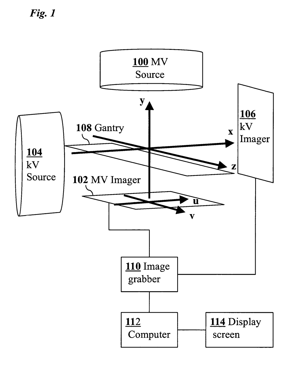
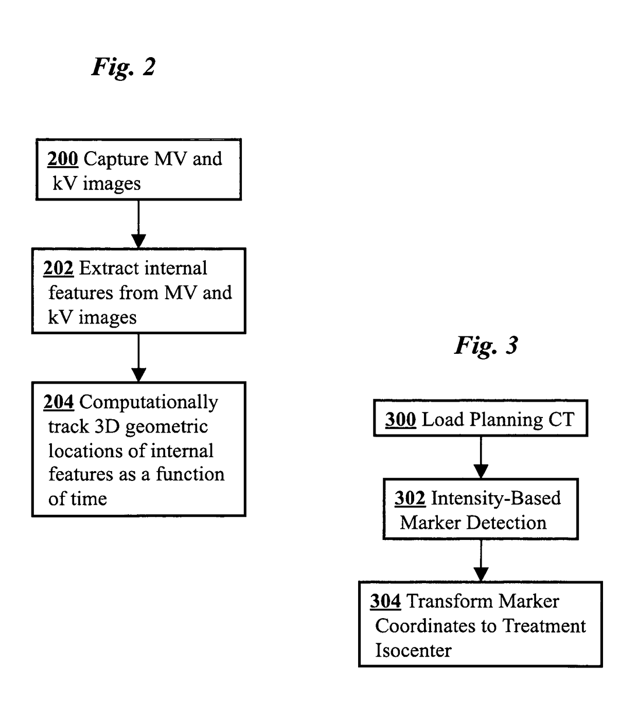
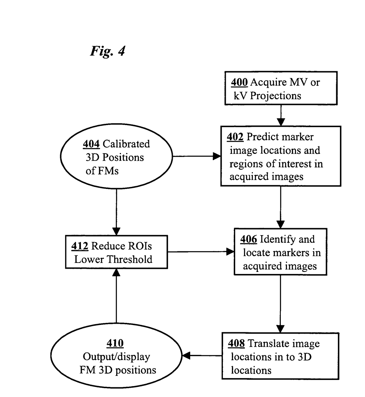
[0015]The present invention provides an improvement of radiotherapy systems together with methods for 3D tumor position monitoring during a treatment delivery. In one aspect, the present invention provides methods to perform 3D internal fiducial or image feature tracking as a function of time based on the combined use of kV and MV imaging. The MV in combination with kV imaging may be used to track the 3D real-time movements of metallic fiducials embedded in a moving subject. Partial geometric and / or temporal information is used to compute the location of fiducials / organs in the event that the MV beam defining aperture blocks the view of the marker / organ on the MV imager during IMRT delivery. The partial information may be used to maintain full 3D tracking as a function of time in the absence of one or both beams (kV & MV) for a short amount of time is also possible.

[0016]A challenge in the use of MV imaging data is that the MV images have low contrast, making it difficult to track i...


PUM


Abstract

A medical imaging-based system and method uses both kV and MV images captured during a treatment period for organ motion tracking. 3D geometric locations of internal features are computationally tracked as a function of time from internal features, such as natural biological features or implanted fiducials, which are computationally extracted from the captured kV and MV images. A partial information method allows 3D tracking to be maintained in the event that imaging information is temporarily not available.

Description

CROSS-REFERENCE TO RELATED APPLICATIONS[0001]This application claims priority from U.S. Provisional Patent Application 61 / 066322 filed Feb. 19, 2008, which is incorporated herein by reference.FEDERALLY-SPONSORED RESEARCH OR DEVELOPMENT[0002]This invention was made with Government support under contract W81XWH-08-1-0128 awarded by the Department of Defense and contract CAI04205 awarded by the National Institutes of Health. The Government has certain rights in this invention.FIELD OF THE INVENTION[0003]The present invention relates generally to medical imaging devices and techniques. More specifically, it relates to methods to accurately monitor the three-dimensional (3D) geometric locations of internal organs as a function of time by the combined use of kilovolt (kV) and megavolt (MV) imaging.BACKGROUND OF THE INVENTION[0004]A goal of x-ray radiotherapy systems is to precisely deliver MV radiation beam to desired locations while minimizing exposure to undesired locations. Movement of...

Claims


Application Information

Patent Timeline
Patent Type & Authority Patents(United States)
IPC IPC(8): G06K9/00
CPCG06T7/2046G06T2207/10081G06T2207/10116G06T2207/30096G06T2207/30204G06T7/251
Inventor WIERSMA, RODNEY D.XING, LEIMAO, WEIHUA
Owner THE BOARD OF TRUSTEES OF THE LELAND STANFORD JUNIOR UNIV
Why Eureka
Try Eureka