

Magnetic resonance method and apparatus for generating a perfusion image
a magnetic resonance and perfusion image technology, applied in the field of magnetic resonance perfusion image generation methods and apparatuses, can solve the problems of inability to immediately obtain final combined perfusion images, and achieve the effect of reducing the effect of motion artifacts and improving the accuracy and quality of the resulting perfusion imag
- Summary
- Abstract
- Description
- Claims
- Application Information
AI Technical Summary
Benefits of technology
Problems solved by technology
Method used
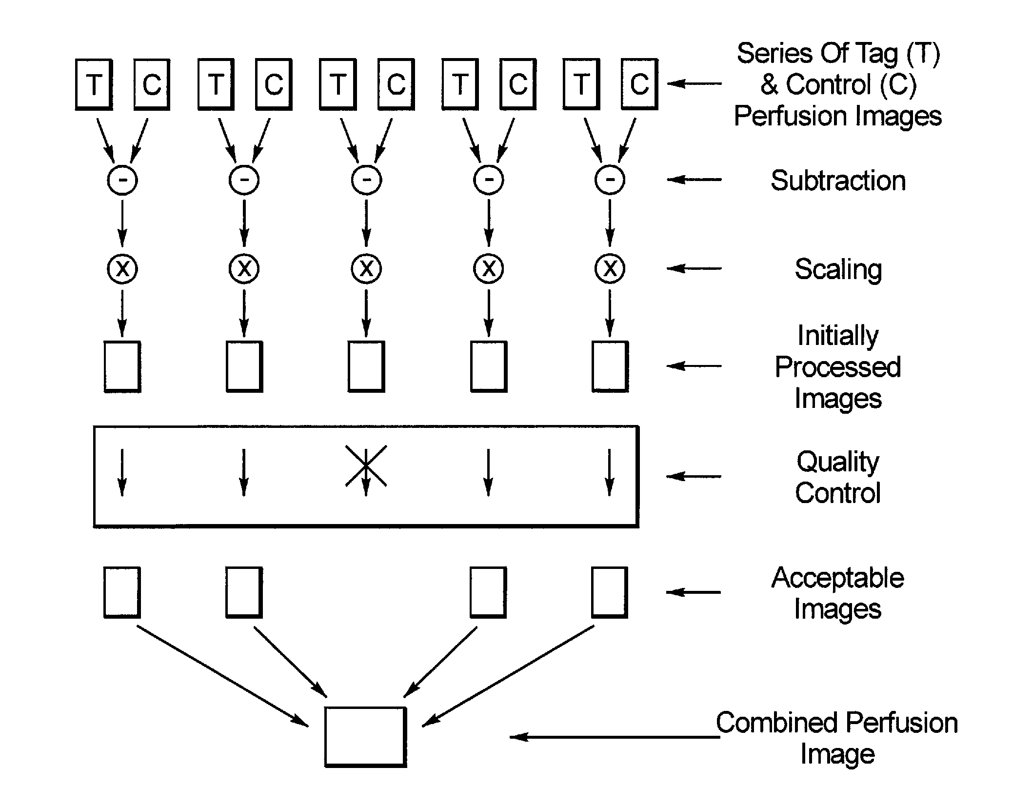
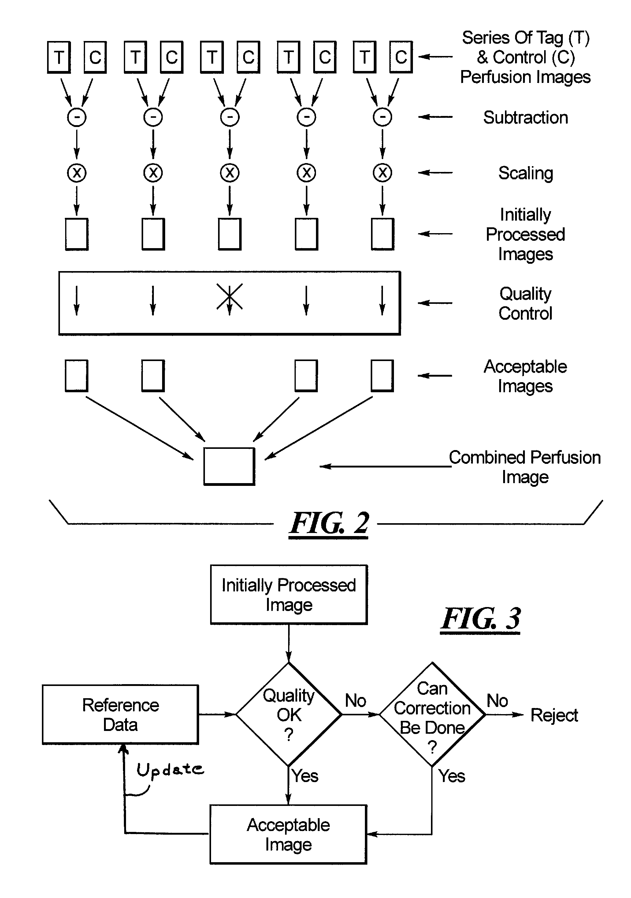
Image

Examples
Embodiment Construction
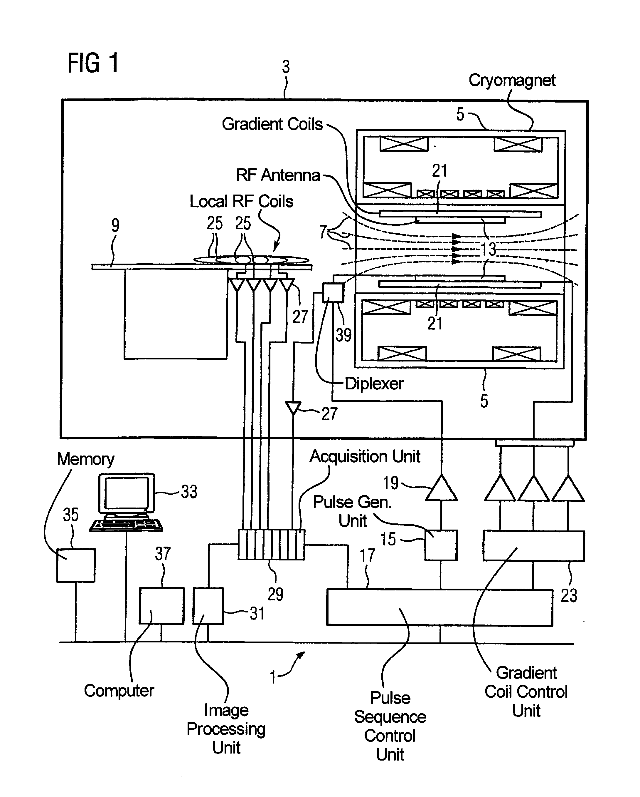
[0017]FIG. 1 schematically shows the design of a magnetic resonance apparatus 1 with its basic components. In order to examine a body by means of magnetic resonance imaging, various magnetic fields tuned to one another as precisely as possible in terms of their temporal and spatial characteristics are applied.
[0018]A strong magnet (typically a cryomagnet 5 with a tunnel-shaped opening) arranged in a radio-frequency shielded measurement chamber 3 generates a static, strong basic magnetic field 7 that typically amounts to 0.2 Tesla to 3 Tesla and more. A body or a body part (not shown here) to be examined is borne on a patient bed 9 and positioned in the homogeneous region of the basic magnetic field 7.
[0019]The excitation of the nuclear spins of the body ensues via magnetic radio-frequency excitation pulses that are radiated via a radio-frequency antenna (shown here as a body coil 13). The radio-frequency excitation pulses are generated by a pulse generation unit 15 that is controlle...
PUM
 Abstract
Description
Claims
Application Information