
Use of eIF-5A to kill multiple myeloma cells
a technology of eif-5a and eif-5a, which is applied in the direction of biocide, drug composition, peptide/protein ingredients, etc., can solve the problems of small decrease in total protein synthesis, many chemotherapy drugs are toxic to actively dividing non-cancerous cells, and lack of protection from ribonucleases, so as to inhibit or slow down the ability of cancer cells to metastasize, reduce tumor growth, and inhibit the effect of tumor growth
Patent Information
- Authority / Receiving Office
- US · United States
- Patent Type
- Applications(United States)
- Current Assignee / Owner
- Publication Date
- 2007-07-05
- Estimated Expiration
- Not applicable · inactive patent
Smart Images
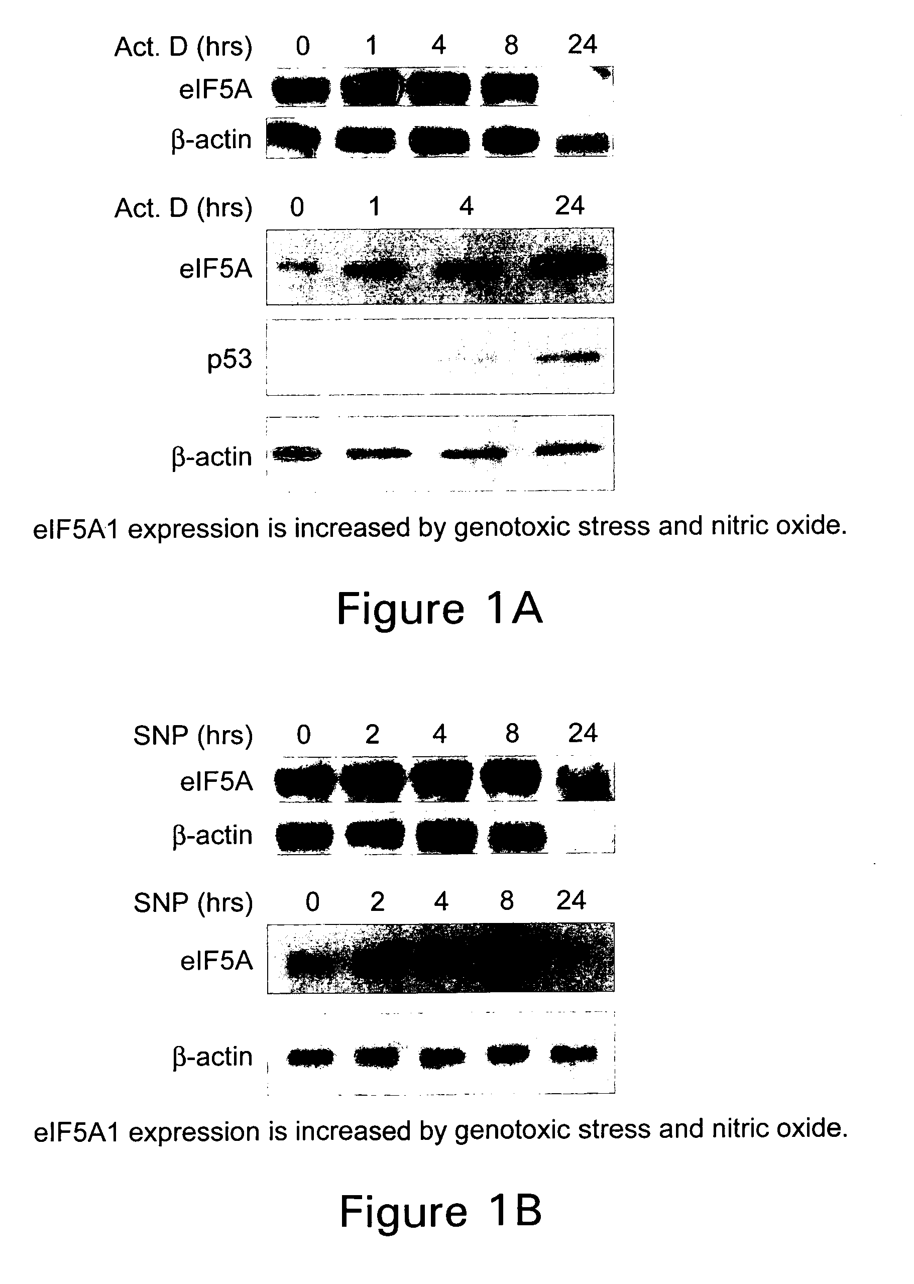
Figure 1 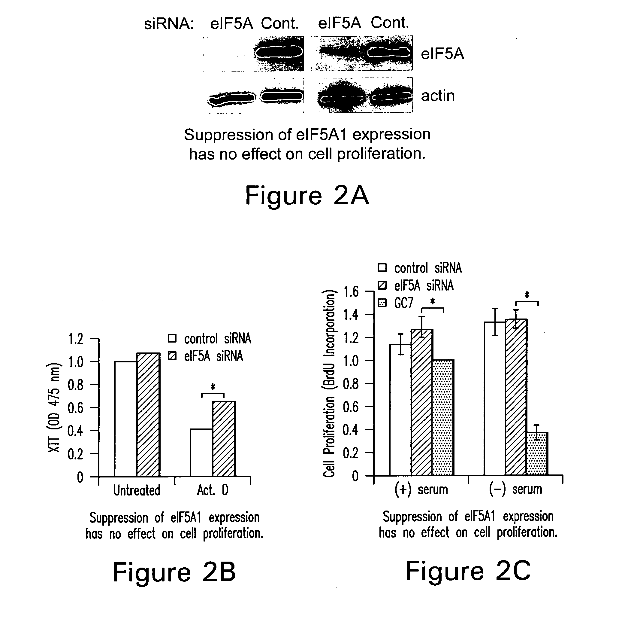
Figure 2 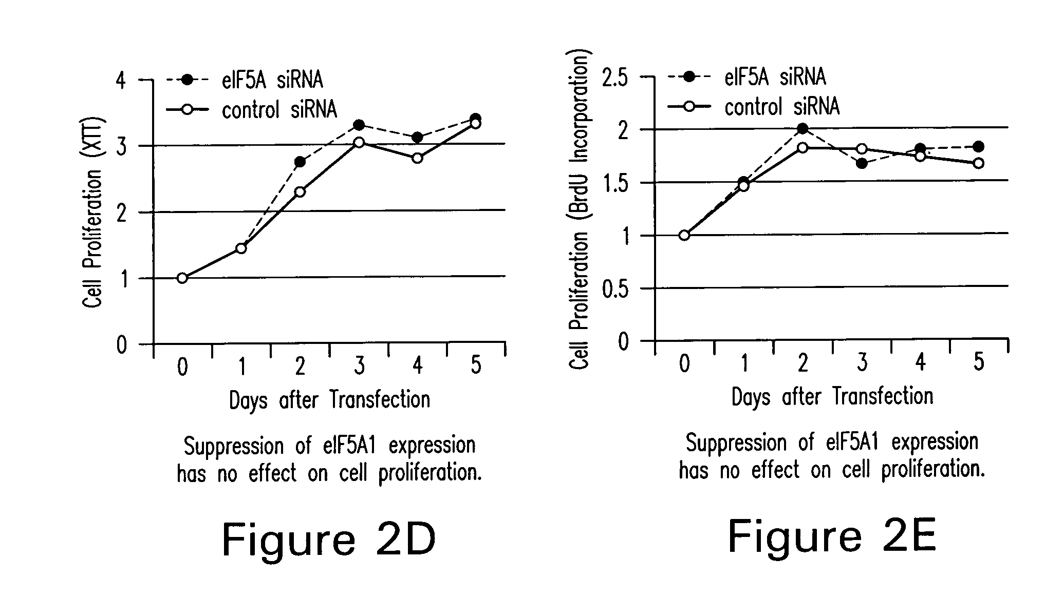
Figure 3
Abstract
Description
RELATED APPLICATIONS
[0001] This application claims priority to U.S. provisional application 60 / 749,604, filed on Dec. 13, 2005 and 60 / 795,168, filed on Apr. 27, 2006, both of which are incorporated by reference in their entirety.FIELD OF THE INVENTION
[0002] The present invention relates to apoptosis-specific eukaryotic initiation factor (“eIF-5A”) and the use of polynucleotides encoding the same to kill multiple myeloma cells, as well as other cancer cells. The present invention relates to the use of apoptosis-specific eIF-5A or referred to as “apoptosis-specific eIF-5A” or “eIF-5A1” as well as the use of the eIF-5A2 isoform to inhibit multiple myeloma, kill multiple myeloma cells, and to inhibit and / or kill other cancer cell growth. BACKGROUND OF THE INVENTION
[0003] Apoptosis is a genetically programmed cellular event that is characterized by well-defined morphological features, such as cell shrinkage, chromatin condensation, nuclear fragmentation, and membrane blebbing. Kerr et...
Examples
example 1
In vitro Experiments
Chemicals
[0096] N1-guanyl-1,7-diaminoheptane (GC7; Biosearch Technologies), an inhibitor of DHS, was used at a concentration of 50 μM. Actinomycin D (Calbiochem) was used at 0.5 or 1.0 μg / ml. Sodium nitroprusside and desferrioxamine were purchased from Sigma and used at a concentration of 3 mM and 500 μM, respectively. Brefeldin A was also acquired from Sigma and used at a concentration of 4 nM.
Cell Culture and Treatment
[0097] The human colon adenocarcinoma cell line, HT-29, was used for cell proliferation and eIF5A localization studies and was a kind gift from Anita Antes (University of Medicine and Dentistry of New Jersey). HT-29 cells were maintained in RPMI 1640 supplemented with 1 mM sodium pyruvate, 10 mM HEPES, and 10% fetal bovine serum (FBS). All other cell lines were obtained from the American Type Culture Collection. CCD112Co is a normal colon fibroblast cell line. RKO is a human colorectal carcinoma cell line (CRL-2577) containing a wild-type p5...
example 2
In vivo Experiments
Mice and Establishment of Tumors
[0112] C57BL / 6 mice were purchased from Charles River, Quebec, Canada at 5-7 weeks of age. Mice were allowed one week to acclimate before experimentation began. B16F10 murine melanoma cells were purchased from ATCC and cultured in DMEM-10% FBS. The cell monolayer was trypsinized and neutralized with MEM-10% FBS. Cells were washed with PBS twice and cell viability was determined by trypan blue staining. For experimental metastasis experiments (Experiments II and III), melanoma tumors were established in the lung by tail vein injection of B16F10 cells into 6-week old mice. B16F10 cells were diluted to 1×10⁶ viable cells / ml in PBS. 200 ul of cells was injected into each mouse via tail vein. For subcutaneous tumor experiments (Experiments IV and V), melanoma tumors were established by subcutaneous injection of 500,000 B16F10 cells into the right flank of 10 to 14-week old mice. At the end of all experiment (when mice became moribund ...
example 3
Experiment II
Construction and Purification of Plasmid DNA
[0113] The pCpG-lacZ expression vector lacking CpG dinucleotides was purchased from InvivoGen, San Diego, USA. An HA-tagged eIF5A1 cDNA was subcloned into the pCpG-lacZ vector by first digesting the plasmid with NcoI and NheI and isolating a 3.1 kb of pCpG vector backbone (thereby removing LacZ coding sequence) and ligating with a PCR amplified cDNA of eIF5A1 containing an HA tag (pCpG-HA5A1). The PCR primers were eIF5A1 for: HA-5A1 for: 5′-GCTCCATGGATGTACCCATACGACGTCCC-3′; and eIF5A1 rev: 5′-CGCGCTAGCCAGTTATTTTGCCATCGCC-3′. The pCpG-lacZ and pCpG-HA5A1 were amplified in E. coli GT115 cultured in LB or 2XYT medium containing 25 μg / ml of zeocin.
[0114] The plasmids were extracted and purified by QIAGEN Endofree Plasmid Giga kit. The DNA concentration was measured by UV absorption at 260 nm and agarose gel electrophoresis.
Tail Vein Injection of Plasmid DNA (Experiment II):
[0115] Plasmid DNA dissolved in 1×PBS (around 200 μ...