
Targeted dual-axes confocal imaging apparatus with vertical scanning capabilities
a confocal imaging and vertical scanning technology, applied in the field of tissue microscopy, can solve the problems of terminal experiments, lower efficiency, distorting pulse profiles, etc., and achieve the effect of improving fluorescence detection efficiency and overcoming blurring
Patent Information
- Authority / Receiving Office
- US · United States
- Patent Type
- Patents(United States)
- Current Assignee / Owner
- Publication Date
- 2018-03-20
Smart Images
Figure 1 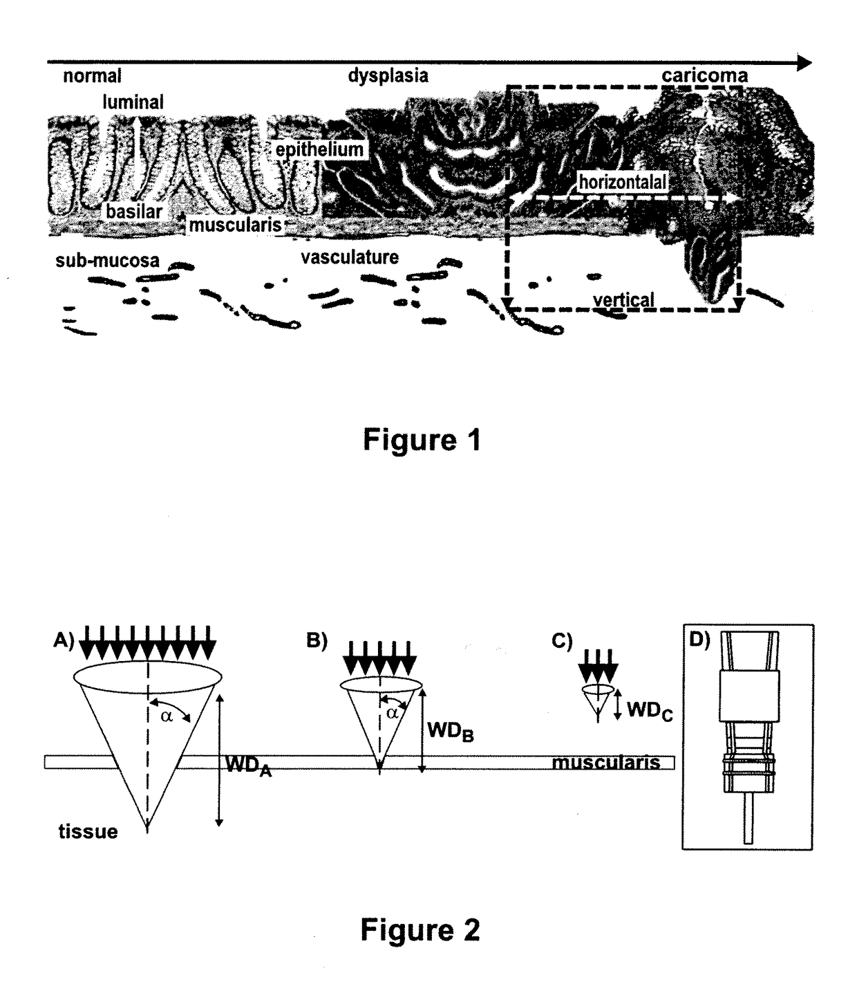
Figure 2 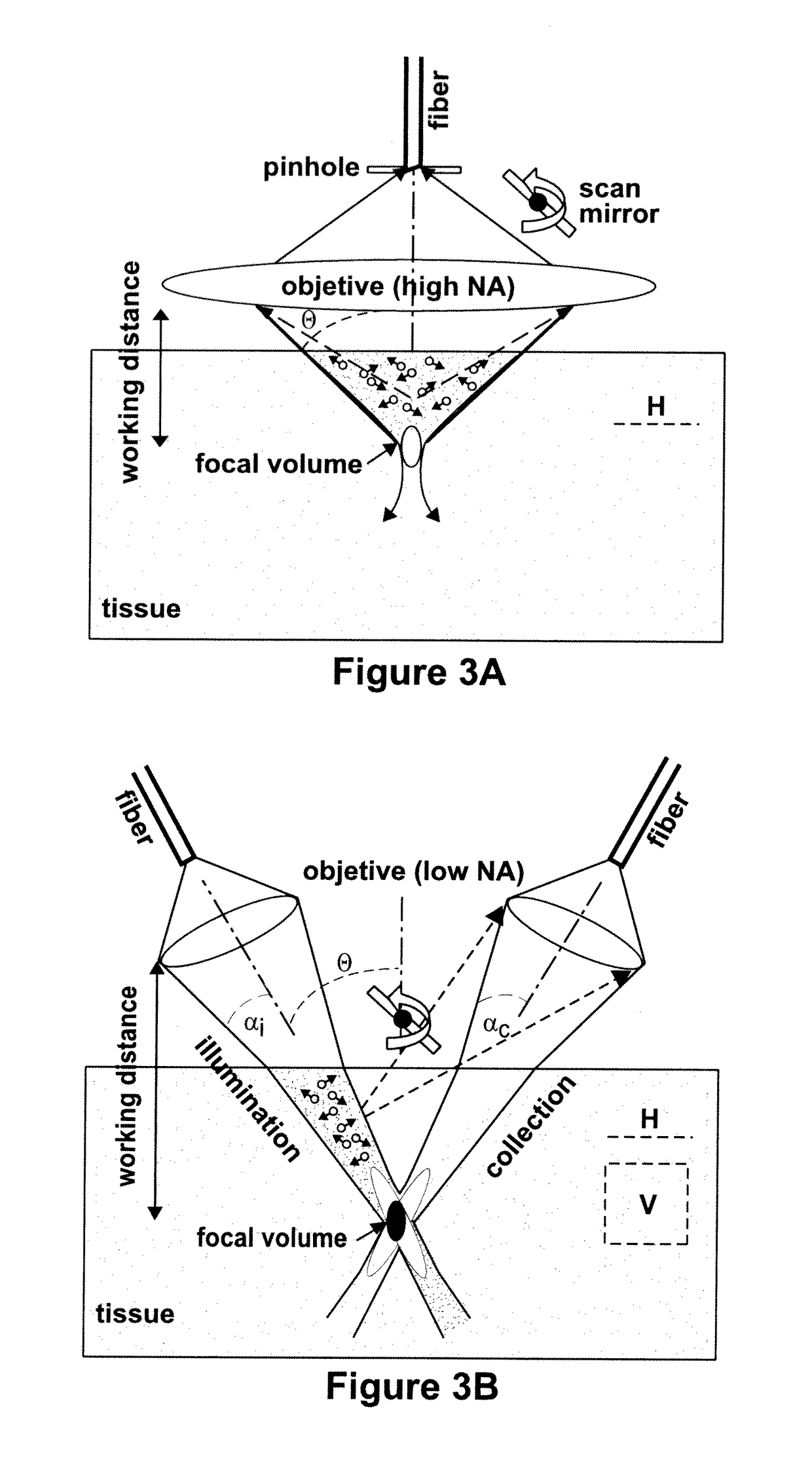
Figure 3
Abstract
Description
CROSS REFERENCE TO RELATED APPLICATIONS
[0001] This application claims the benefit of U.S. Provisional Application No. 61 / 256,937, filed Oct. 30, 2009, the entirety of which is expressly incorporated herein by reference.STATEMENT OF GOVERNMENT INTEREST
[0002] This invention was made with government support under CA136429 and CA093990 awarded by the National Institutes of Health. The government has certain rights in the invention.BACKGROUND OF THE DISCLOSURE
[0003] Field of the Disclosure
[0004] The disclosure relates generally to microscope imaging of tissue and, more particularly, to devices for dual-axes confocal microscopy.
[0005] Brief Description of Related Technology
[0006] While tremendous advances have been made in technologies for whole body molecular imaging, including PET, SPECT, MRI, and ultrasound, techniques for visualizing biological phenomenon with sub-cellular resolution in optically thick tissue are lacking. In addition, these conventional techniques lack the ability to obtain...
Examples
example 1
[0129]Peptides have demonstrated tremendous potential for use as molecular probes to identify cell surface targets in vivo. In addition to high clonal diversity, small size, low immunogenicity and compatibility with fluorescence dyes, peptides exhibit rapid binding kinetics and high binding specificity. Targeting peptides have been developed using techniques of phage display, a powerful combinatorial method that uses recombinant DNA technology to generate a complex library of peptides for selection by preferential binding to cell surface targets. The protein coat of bacteriophage, such as the filamentous M13 or T7, is genetically engineered to express a very large number (>109) of different peptides with unique sequences to achieve high affinity binding (New England Biolabs, Inc. Ph.D.7™ phage display peptide library kit. Instruction manual 2006; ver 2.7). Selection is then performed by biopanning the phage library against cultured cells that overexpress the target (Zurita et al., C...
example 2
[0132]Small animal models play an important role in the study of colon cancer biology because the natural history of these tumors develop over an abbreviated time course (between 6 to 12 months) compared to that of humans.
[0133]A genetically engineered mouse model has been developed that spontaneously expresses polyps in the distal colon and rectum (Hinoi et al., Cancer Res. 2007; 67:9721-30; Akyol et al., Nat Methods 2008; 5:231-3). This small animal model can be used to demonstrate the imaging performance of the novel dual-axes confocal microscope and to evaluate affinity binding of the fluorescence-labeled peptides. The model is based on mutations in the adenomatous polyposis coli (APC) gene to initiate cancer development in the distal colon. Previous mouse models based on the APC gene expressed tumor mainly in the small intestine where it would be extremely difficult to access using techniques of intra-vital microscopy in a non-terminal experiment. A new mouse model of colon can...
example 3
[0138]Chemokine receptor CXCR7 was recently identified as a highly promising new molecular target of angiogenesis in breast cancer.
[0139]On immunohistochemistry, 97% of human primary breast tumors expressed CXCR7 in the tumor vascular endothelium, while the receptor was undetectable in normal breast tissue, as shown in FIG. 26C (Miao et al., Proc Natl Acad Sci USA 2007; 104:15735-15740). CXCR7 expression in tumor vasculature can be demonstrated on immunohistochemistry using the mouse monoclonal antibody 11G8 (Burns et al., J Exp Med 2006; 203:2201-2213). Invasive ductal carcinoma of the breast reveals CXCR7 expression in the tumor vasculature (arrow), magnification 10×, shown in FIG. 26A. Endothelial staining of a blood vessel (arrow) from a) is shown in FIG. 26B, magnification 40×. Normal breast tissue vasculature shows no CXCR7 expression, as shown in FIG. 26C, magnification 10×. CXCR7 negative blood vessels from c) are shown in FIG. 26D, magnification 40×. In addition, CXCR7 imag...