Ultrasonic joint probe used in blood vessel

An ultrasonic detection and blood vessel technology, applied in the field of medical devices, can solve the problem that the probe cannot be accurately focused, and achieve low cost effects

Active Publication Date: 2017-12-19
THE FIRST AFFILIATED HOSPITAL OF THIRD MILITARY MEDICAL UNIVERSITY OF PLA
View PDF9 Cites 0 Cited by
  • Summary
  • Abstract
  • Description
  • Claims
  • Application Information

AI Technical Summary

Problems solved by technology

[0004] The present invention intends to provide an ultrasonic combined probe for use in blood vessels to solve the problem that the existing probe cannot be accurately focused on the target nerve

Method used


Image

  • Ultrasonic joint probe used in blood vessel

Examples


Embodiment Construction

[0027] The present invention will be described in further detail below by means of specific embodiments:

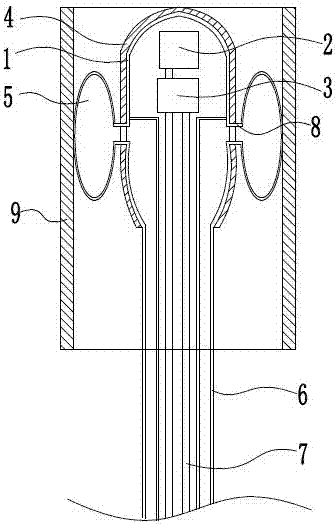
[0028] The reference signs in the drawings of the description include: probe body 1 , ultrasonic probe 2 , high-energy focusing head 3 , main sleeve 4 , balloon 5 , catheter 6 , guide wire 7 , gas-filled tube 8 , and blood vessel 9 .

[0029] The embodiment is basically as attached figure 1 Shown: the ultrasonic joint probe used in the blood vessel 9, including the probe body 1 for extending into the blood vessel 9 and the catheter 6 communicating with the probe body 1 for extending out of the body; the probe body 1 is provided with an ultrasonic probe 2 and A high-energy focusing head 3; a main tube sleeve 4 connected to the probe body 1 is arranged outside the probe body 1; the main tube sleeve 4 is a shuttle-shaped structure with small ends and a large middle. The front finger is close to the end extending into the blood vessel 9 , and the back finger is close to the ...


PUM


Abstract

The invention discloses an ultrasonic joint probe used in the blood vessel. The ultrasonic joint probe comprises a probe body extending into the blood vessel and a catheter communicated with the probe body and extending out of the body, wherein an ultrasonic detection head and a high-energy focusing head are arranged in the probe body, a main tube casing communicated with the probe body is arranged outside the probe body, three air bags are arranged on the main tube casing, the catheter is provided with a middle interlayer, and a channel for a wire to pass through is formed in the catheter. The middle interlayer of the catheter is used for inflating the air bags. The ultrasonic joint probe can be accurately focused on the target spot nerve.

Description

technical field [0001] The invention relates to the field of medical devices, in particular to an ultrasonic combined probe used in blood vessels. Background technique [0002] High-intensity focused ultrasound (HIFU) is a kind of energy that can be precisely focused and non-contact damage. Through intravascular intervention, it can be sent to the position of the target nerve to release heat energy for nerve damage treatment. For the treatment of cardiac pre-excitation syndrome and atrial fibrillation. At present, the independent balloon ultrasound probe cannot accurately determine the specific position of the target nerve in the blood vessel, and cannot accurately locate the focal point of the probe on the target nerve trunk or ganglion. In addition, because the balloon of the balloon ultrasound probe is a cylindrical or ellipsoid-like structure, it only has two modes of fixation in the blood vessel, floating and fixed, and the balloon probe cannot adjust the distance to t...

Claims


Application Information

Patent Timeline
IPC IPC(8): A61B8/12A61B8/08A61N7/02
CPCA61B8/0891A61B8/12A61B8/445A61N7/02A61N2007/0021
Inventor 崔剑肖航赵敬华
Owner THE FIRST AFFILIATED HOSPITAL OF THIRD MILITARY MEDICAL UNIVERSITY OF PLA
Why Eureka
Try Eureka