Immunohistochemical pathological image CD3 positive cell nucleus segmentation method and system

A pathological image and immunohistochemical technology, applied in the field of image processing, can solve the problem of spending a lot of time and energy, and achieve the effect of improving precision and accuracy

Active Publication Date: 2019-11-05
GUANGDONG GENERAL HOSPITAL
View PDF2 Cites 10 Cited by
  • Summary
  • Abstract
  • Description
  • Claims
  • Application Information

AI Technical Summary

Problems solved by technology

At present, it takes a lot of time and energy for medical staff to segment, label and count CD3 cells in pathological image...

Method used


Image

  • Immunohistochemical pathological image CD3 positive cell nucleus segmentation method and system
  • Immunohistochemical pathological image CD3 positive cell nucleus segmentation method and system
  • Immunohistochemical pathological image CD3 positive cell nucleus segmentation method and system

Examples


Embodiment

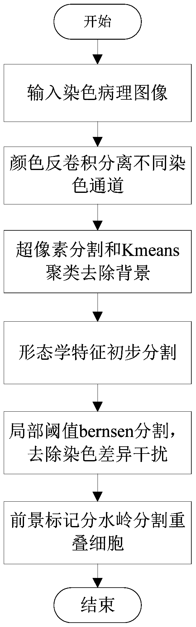
[0047] Such as figure 1 As shown, this embodiment provides a method for segmenting CD3-positive cell nuclei in immunohistochemical pathological images, comprising the following steps:

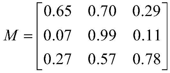
[0048] S1: Color deconvolution is performed on the original RGB-encoded immunohistochemical pathological image, and the two staining channels of hematoxylin (Haematoxylin, H) and diaminobenzidine (3,3'-Diaminobenzidine, DAB) are separated, and only DAB is used Stained CD3-positive cells were segmented;

[0049]In this embodiment, the color deconvolution algorithm is based on the color information acquired by the RGB camera, and based on the specific absorption of the RGB component light of the stain used in the immunohistochemical technique, the effect of each stain on the image is calculated separately, and the deconvolution The product refers to the process of calculating the unknown input with the known output and partial input, where the output is the CD3 staining map, the known input is H...


PUM


Abstract

The invention discloses an immunohistochemical pathological image CD3 positive cell nucleus segmentation method and a system, and the method comprises the steps: carrying out the color deconvolution of an immunohistochemical pathological image, and separating a staining channel; segmenting the image into irregular pixel blocks by adopting superpixels, carrying out kmeans clustering to distinguishan image background, and removing the image background; performing image segmentation based on the morphological characteristics to obtain a preliminarily segmented cell nucleus first region image L1and a to-be-processed first image C1; performing local threshold bernsen segmentation and morphological feature segmentation on the first image C1 to be processed to obtain a segmented cell nucleus second region image L2 and a second image C2 to be processed; performing foreground marking on the second image C2 to be processed, and segmenting a third region image L3 of the cell nucleus by adoptinga watershed algorithm; and forming a cell nucleus segmentation image by the cell nucleus first region image L1, the cell nucleus second region image L2 and the cell nucleus third region image L3. Themethod is high in robustness and accurate in segmentation, and can meet the requirements of practical application.

Description

technical field [0001] The invention relates to the technical field of image processing, in particular to a method and system for segmenting CD3-positive cell nuclei in immunohistochemical pathological images. Background technique [0002] CD3 is an important leukocyte differentiation antigen that exists on the surface of almost all T cells and is a membrane antigen that constitutes the T cell antigen receptor (TCR). When an antigen binds to TCR, it participates in the transmission of the signal to the cell, and is an important membrane antigen involved in the discovery of various T cell functions. At present, it takes a lot of time and energy for medical staff to segment, label and count CD3 cells on pathological images. Therefore, a more accurate means of cell segmentation in pathological images is needed to reduce the pressure on medical staff. Contents of the invention [0003] In order to reduce the burden on medical staff and realize automatic segmentation of CD3 ce...

Claims


Application Information

Patent Timeline
IPC IPC(8): G06T7/11G06T7/136G06T7/194
CPCG06T2207/10056G06T7/11G06T7/136G06T7/194
Inventor 刘再毅梁长虹覃杰赵可王瑛陈鑫黄燕琪姚溯李振辉
Owner GUANGDONG GENERAL HOSPITAL
Why Eureka
Try Eureka