Gumnosperm pollen tube microfilament framework fluorescent marking method and its uses

A fluorescent labeling, gymnosperm technology, applied in plant cells and other directions, can solve problems such as the lag of biological research, and achieve the effects of good labeling effect, high application value and simple operation.

Inactive Publication Date: 2006-12-27
INST OF BOTANY CHINESE ACAD OF SCI
View PDF0 Cites 8 Cited by
  • Summary
  • Abstract
  • Description
  • Claims
  • Application Information

AI Technical Summary

Problems solved by technology

So far, no effective methods and techniques for labeling microfilament skeletons in gymnosperm pollen grains and pollen tubes have been reported, resulting in a lag in biological research related to gymnosperms compared to angiosperms

Method used


Image

  • Gumnosperm pollen tube microfilament framework fluorescent marking method and its uses
  • Gumnosperm pollen tube microfilament framework fluorescent marking method and its uses

Examples


Embodiment 1

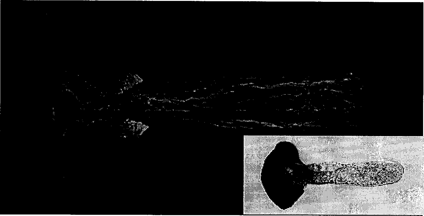
[0025] Example 1. Fluorescent labeling of the microfilament skeleton of white rod pollen tubes and its microscopic observation

[0026] Firstly, the method of the present invention is used to carry out fluorescent labeling to the microfilament skeleton of the white pole pollen tube, comprising the following steps:

[0027] 1) Collect the white stem pollen tube samples to be marked, and prepare them in freshly prepared 50mM Pipes buffer (50mM PIPES, 0.5mM MgCl) containing 3% paraformaldehyde 2 , 1mM EGTA, pH 6.9) and quickly pumped and fixed for 30min;

[0028] 2) Wash 3 times with 50mM Pipes buffer to remove residual paraformaldehyde;

[0029] 3) Put the pollen tubes in 0.25% glycerol and infiltrate at room temperature for 40 minutes;

[0030] 4) wash 3 times with 50mM Pipes buffer;

[0031] 5) Incubate the pollen tube with 200 nM TRITC-phalloidin (Sigma, USA) in the dark at room temperature for 1 h. Then, drop into 50% glycerol containing 0.2% Propyl gallate (Sigma Compan...

Embodiment 2

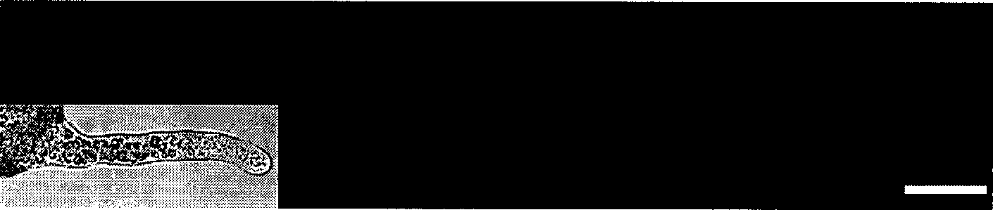
[0032] Example 2. Fluorescence labeling and microscope observation of the microfilament skeleton of Pine pine pollen tube

[0033] Firstly, the method of the present invention carries out fluorescent labeling to the pine pollen tube microfilament skeleton, comprising the following steps:

[0034] 1) Collect the white bark pine pollen tube samples to be marked, and prepare them in freshly prepared 60mM Pipes buffer (60mM PIPES, 0.5mM MgCl) containing 4% paraformaldehyde 2 , 1mM EGTA, pH 6.9) in fast pumping and fixation for 50min;

[0035] 2) Washing 4 times with 60mM Pipes buffer to remove residual paraformaldehyde;

[0036] 3) Put the pollen tubes in 0.2% glycerin and infiltrate at room temperature for 60 minutes;

[0037] 4) wash 4 times with 60mM Pipes buffer;

[0038] 5) Incubate the pollen tubes with 250 nM TRITC-phalloidin in the dark at room temperature for 1.5 h. Then, drop in 60% glycerin containing 0.2% Propyl gallate to the fluorescently labeled pollen tube samp...


PUM


Abstract

The invention discloses a method for fluorescence labeling of gymnosperm pollen tube fibril bone and its application and is to provide a method for fluorescence labeling of gymnosperm pollen tube fibril bone and the application of this method in microscope inspection of gymnosperm pollen tube fibril bone. The said labelling method includes the following steps: 1) fixing the gymnosperm pollen tube with 40-60mM Pipes cushion solution containing 2-4 % cavaform; 2) washing the pollen tube; 3) placing the pollen tube in 0.2-0.3 % glycerin to infiltrate for 30-60min; 4) washing the pollen tube; 5) dyeing the pollen tube and avoiding the sun with 150-250nM phalloidine by fluorescence labeling for 0.5-1.5h and fluorescence labeling the fibril bone. The invention is of simple operation and is quick, has small damage to the cells, and has good labeling effect, which will play an important role in study gymnosperm pollen tube cell bone and have a high actual application value.

Description

technical field [0001] The present invention relates to a fluorescent labeling method of pollen tube microfilament skeleton in the field of plant biotechnology and its application, in particular to a method for fluorescent labeling of gymnosperm pollen tube microfilament skeleton and the use of the method for gymnosperm pollen pollen tube microfilament Microscopic observation of silk skeletons. Background technique [0002] As the carrier of the male reproductive unit in the fertilization process of flowering plants, pollen tube has typical apical growth characteristics, so it has become an ideal model system for studying the growth of cell polarity in the field of biotechnology. In addition, the pollen tube system is also of great significance to the study of the interaction between cells and the study of signal transduction. The cytoskeleton is the basic organizational structure of the pollen tube, and it participates in important physiological processes such as cell shap...

Claims


Application Information

Patent Timeline
Patent Type & Authority Applications(China)
IPC IPC(8): C12N5/04
Inventor 林金星陈彤王玉花
Owner INST OF BOTANY CHINESE ACAD OF SCI
Why Eureka
Try Eureka