Implant for correcting skeletal mechanics

a technology applied in the field of medical devices for enhancing and correcting skeletal mechanics, can solve the problems of no longer being able to support the first metatarsal, excessive strain on the soft tissues supporting this bone, and stretching of the soft tissues

Inactive Publication Date: 2009-09-03
GRAHAM MICHAEL
View PDF9 Cites 72 Cited by
  • Summary
  • Abstract
  • Description
  • Claims
  • Application Information

AI Technical Summary

Problems solved by technology

Excessive pronation (hyperpronation) of the foot leads to abnormal motion to the first metatarsal resulting in excessive strain on the soft tissues supporting this bone.
After a prolonged period of these excessive forces the soft tissues will stretch out and no longer be able to support the first metatarsal.
This instability leads to an abnormal deviation of the first metatarsal bone resulting in foot pathology.
The end result is that there will be instability at both the proximal and distal joints of the first metatarsal bone.
This deformation causes instability of the first metatarsal bone and contributes to the medial shift of the metatarsal bone.
The problem with non-surgical treatment options is that it is ineffective in eliminating the causative factor, meanwhile every step leads to further deformity of the metatarsal bone.
The problem with this deformity is intrinsic so external remedies are ineffective in controlling the deforming forces.
These osseous procedures of the first metatarsal bone only provide for a cosmetic effect while the instability of the first metatarsal / first cuneiform joint still exists.
These types of procedures straighten the metatarsal bone with respect to the hallux but leave instability at the first metatarsocuneiform joint.
Since the instability at the first metatarsocuneiform joint still exists, the first metatarsal bone will eventually deviate again and lead, again, to the occurrence of the overall deformity.
These procedures lead to a long recovery periods, at least six months, and can fail.
Unfortunately, this transfers the body's weight to the second metatarsal head instead of the first metatarsal head.
Often, further pathology ensues leading to callus formation under the ball of the foot leading to further pain, and possible ulceration.
The problem with these plates is that they are use to stabilize the fusion site between these two bones and overall minic a screw or staple.
There is still a shortening of the first metatarsal bone leading to the possibility of other ill effects.
These plates are rather bulky and usually had to be removed after the arthrodesis of the two bones was achieved.

Method used


Image

  • Implant for correcting skeletal mechanics
  • Implant for correcting skeletal mechanics
  • Implant for correcting skeletal mechanics

Examples


Embodiment Construction

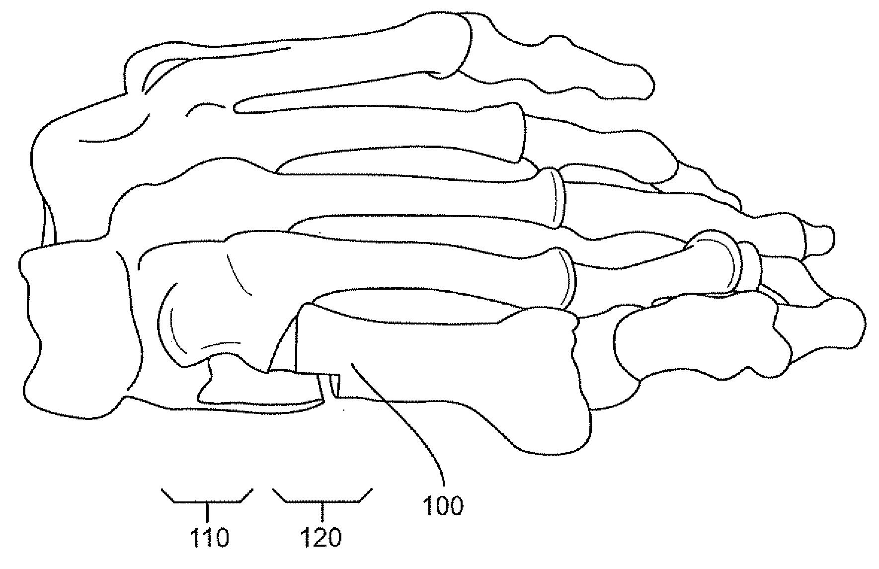
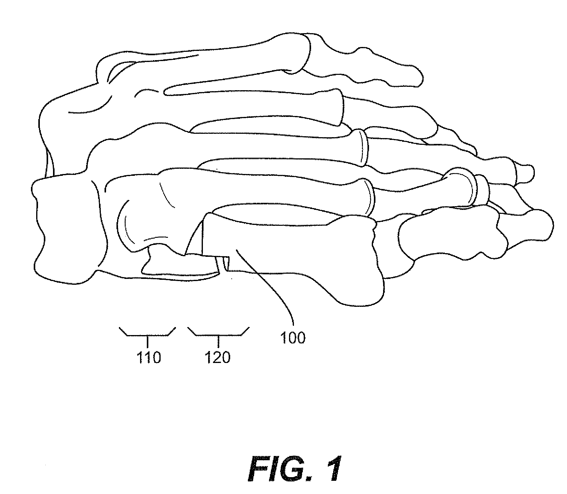
[0019]Turning to figure three, the plate has a proximal (back portion) and distal (front portion) end. The proximal wider end of the plate has two screw recesses 354, 355 for fixation of the plate in the first cuneiform. The wedge on the lateral side of the plate will be introduced between the first metatarsocuneiform joint to correct transfer of weight from the first cuneiform to the base of the first metatarsal bone. The distal narrower end of the plate also has two screw recesses 356, 357 for fixation onto the first metatarsal bone. Also found on the plate are two holes, one the proximal 352 and distal 350 segment for the first metatarsal and the first cuneiform bones. These holes are for temporary pin stabilization to hold the plate in place while the screws placed in the screw recesses (356, 357, 352, 350) to fixate the plate to the bones. See also similar screw recesses in FIGS. 4 and 5 (456, 457, 452, 450; 556, 557, 552, 550).

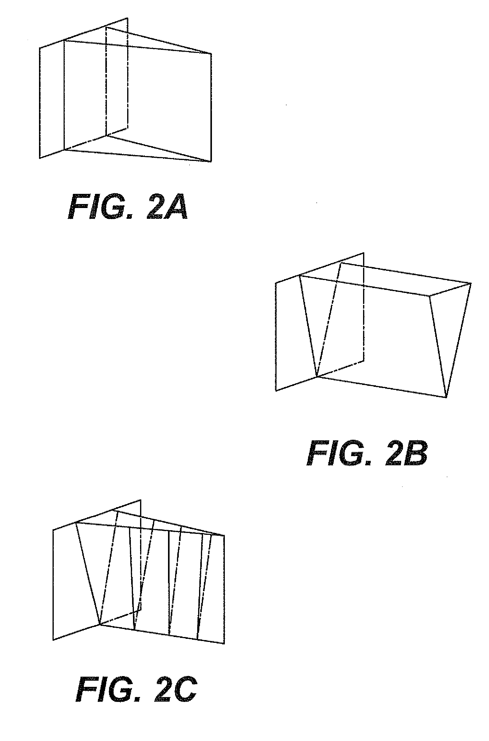
[0020]The interpositioned wedge will consist of va...


PUM


Abstract

Deformities present on the end of a bone, for example the end of the metatarsal bone making up part of the metatarsocuneiform joint, can lead to deformities such as bunions. These deformities are treatable with an implant that comprises a plate with a wedge extending perpendicular from the plate. Following removal of cartilage from the joint, deformed portions at the end of the bone are removed and the wedge is inserted in the joint and held in place when the plate is attached to the bones flanking the joint. This effectively fuses the two bones together. The wedge can be shaped in various ways depending on the particular deformity present.

Description

BACKGROUND OF THE INVENTION[0001]This invention relates to a medical apparatus for enhancing and for correcting skeletal mechanics. More specifically, this invention relates to the correction of certain bone alignment deformities that impair optimal biped mechanics of the foot.[0002]Excessive pronation (hyperpronation) of the foot leads to abnormal motion to the first metatarsal resulting in excessive strain on the soft tissues supporting this bone. After a prolonged period of these excessive forces the soft tissues will stretch out and no longer be able to support the first metatarsal. This instability leads to an abnormal deviation of the first metatarsal bone resulting in foot pathology. The deforming forces acting on the first metatarsal leads to three possible deviations: (1) pure medial deviation of the metatarsal, (2) dorsiflexion of the metatarsal, and (3) the combination of the previous two, dorsomedial deviation. When the first metatarsal deviates medially (toward the body...

Claims


Application Information

Patent Timeline
Patent Type & Authority Applications(United States)
IPC IPC(8): A61B17/58
CPCA61B17/809A61B17/8061
Inventor GRAHAM, MICHAEL
Owner GRAHAM MICHAEL
Why Eureka
Try Eureka